Key takeaway: Researchers found lymph node–like immune hubs in mouse skull bone marrow that contain B cells and T follicular helper–like cells and appear to take part in CNS antigen surveillance. In mouse glioma models, disrupting these hubs worsened tumor growth and survival, while a local immune-boosting gel improved outcomes. Similar immune cells were observed in human skull samples, but the organized structures and therapeutic potential in people remain unconfirmed. The discovery reframes brain–immune interactions but is not yet a human treatment.
Hidden Immune Hubs in the Skull May Act as a First Line of Defense Against Brain Tumors
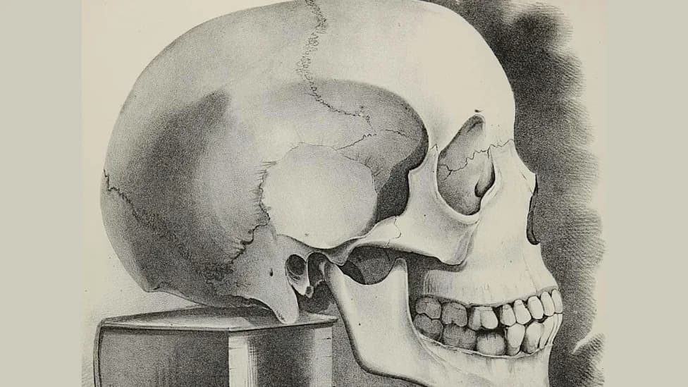
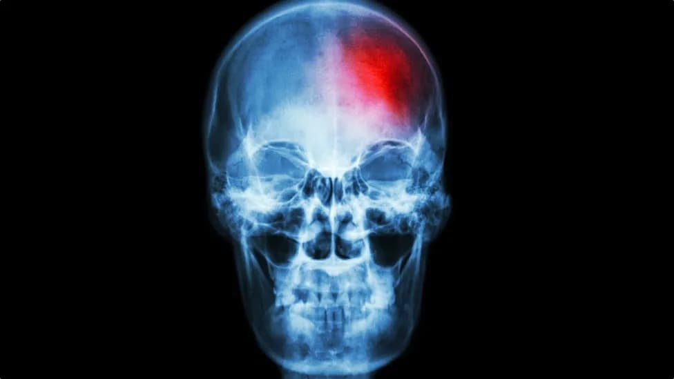
Human Skull
For most of human history the skull has been thought of primarily as a protective shell for the brain. New research from Washington University School of Medicine in St. Louis suggests the skull may also house organized immune hubs that help monitor and respond to threats near the central nervous system — at least in mice.
What the Study Found
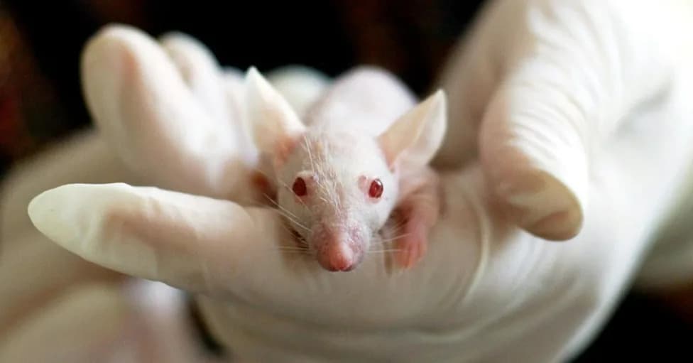
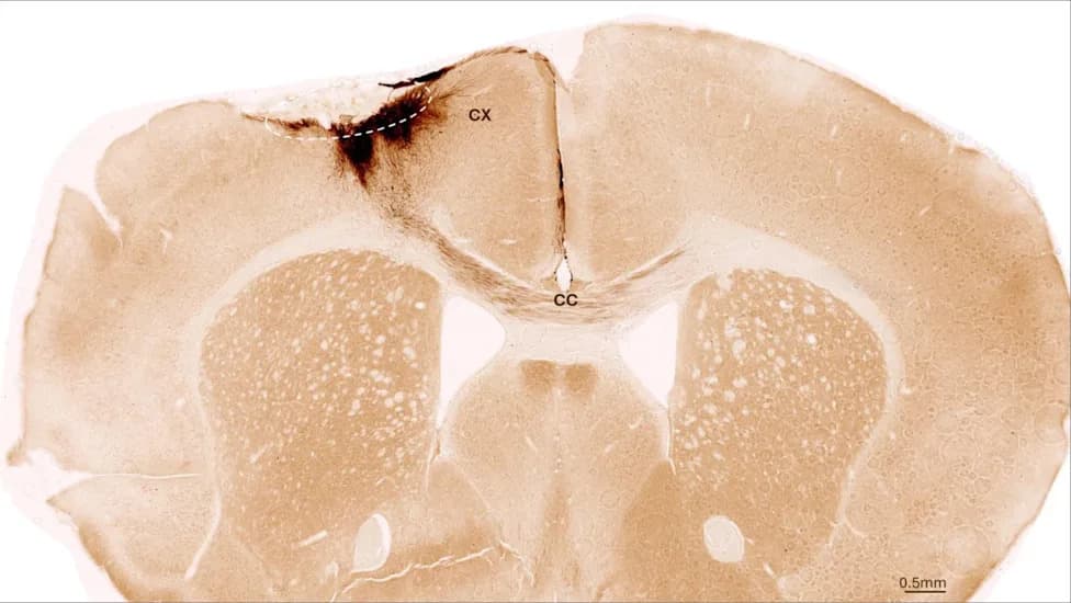
The team identified lymph node–like structures in mouse skull bone marrow that contain B cells and T follicular helper–like (Tfh-like) cells, immune cells that coordinate adaptive responses. The study, titled "Functional Role of Skull Lymphoid Structures in CNS Immunosurveillance," reports these formations appear to participate in antigen surveillance for the central nervous system and to shape anti-tumor immunity in mouse models.
Evidence From Mouse Experiments
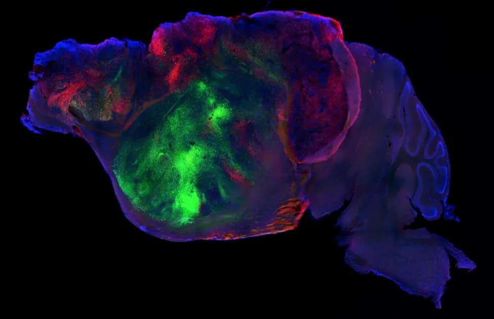
Functional tests in glioma and glioblastoma mouse models showed that disrupting these skull immune structures allowed tumors to grow faster and reduced animal survival. Conversely, a localized treatment — a gel containing three immune-stimulating proteins applied beneath the scalp — boosted anti-tumor responses and improved survival in those mice. These results support a role for the skull-associated lymphoid formations in coordinating local immune defenses in animal models.
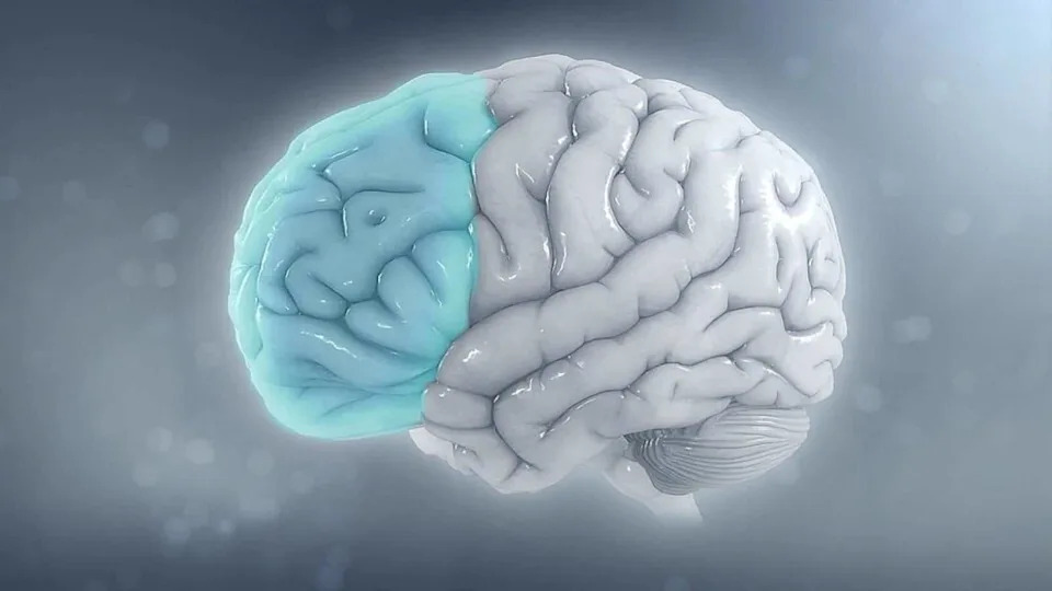
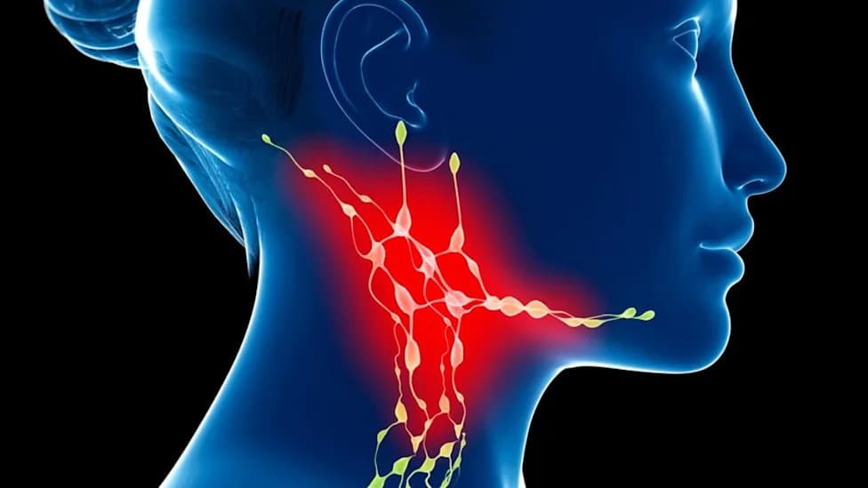
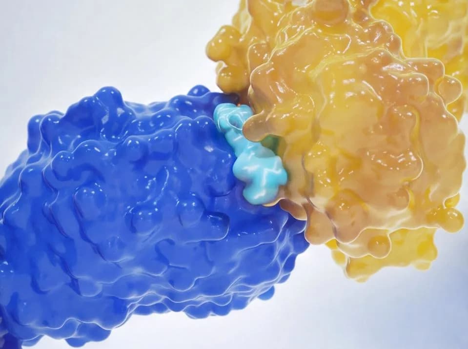
Image Credit:https://www.scientificanimations.com/,CC BY-SA 4.0, via Wikimedia Commons
Human Relevance and Limits
Researchers detected similar immune cell types in human skull bone marrow samples, but they have not yet demonstrated the same organized, lymph node–like structures or confirmed equivalent function in people. The authors caution that findings in mice provide important clues but do not directly translate into human therapies. The discovery is best described as identification of a previously unrecognized immune structure in mice with potential implications for human biology, not as proof of a new human organ or an established clinical treatment.
Context and Significance
This work builds on prior discoveries — such as dural lymphatic vessels and channels linking skull marrow to nearby tissues — that have shifted how scientists view brain–immune interactions. Rather than an immune-isolated organ, the brain and its surrounding tissues appear to participate in active immune communication. The new findings change where researchers may look for mechanisms that control brain inflammation and tumor immunity.
Jonathan Kipnis, PhD (senior author): "This study reveals that the skull bone marrow is far more than just a structural framework. It harbors previously unrecognized hubs for brain-specific immune responses."
Jang Hyun Park, PhD (first author): "We have never seen such structures in healthy bone marrow before."
Bottom line: The study reveals an unexpected immune role for skull bone marrow in mice and highlights a promising direction for future research into brain immunity and cancer — but it does not establish a human therapy or a newly classified human organ.
Image Credit: https://www.scientificanimations.com/, CC BY-SA 4.0, via Wikimedia Commons
Help us improve.