Stanford researchers implanted lab-grown human neural tissue into mice engineered to lack a cortex and hippocampus, resulting in grafts that in some animals filled up to half the brain by volume. The transplanted tissue vascularized and produced around four million human neurons that formed some connections with mouse circuitry but remained immature (mid-gestation). The model revealed striking vulnerability of human neurons to prolonged oxygen deprivation and produced von Economo neurons relevant to frontotemporal dementia research. The work proceeded under stated ethical oversight, though bioethicists urge continued monitoring and debate.
Stanford Grows Human Neural Tissue That Replaces Up to Half A Mouse Brain — A New Model For Studying Brain Disorders
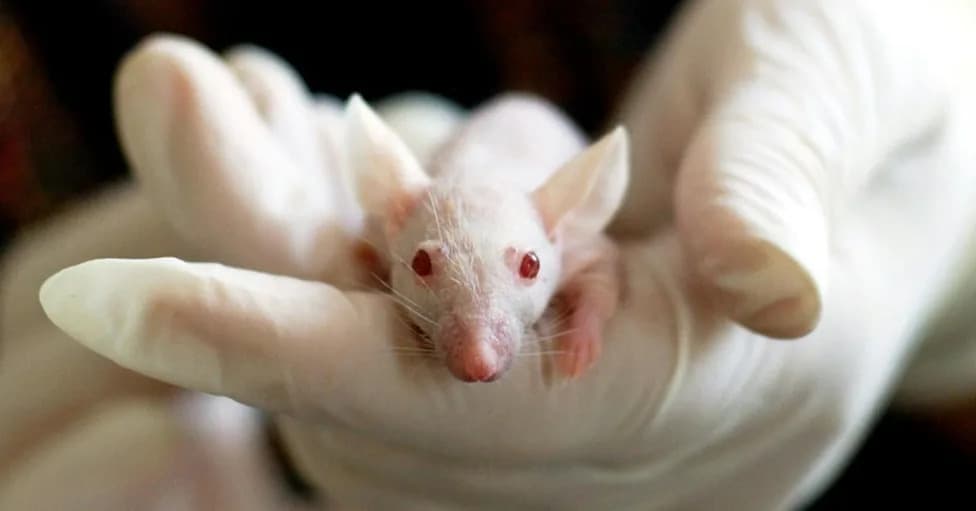
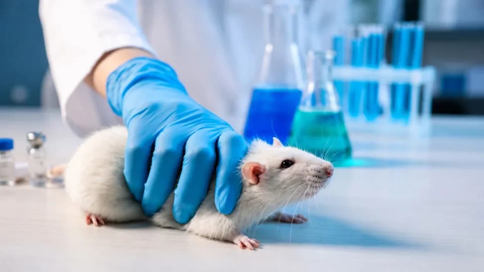
White lab mouse
Researchers at Stanford have implanted lab-grown human neural tissue into specially engineered mice, and in some animals the human grafts now occupy roughly half the brain’s volume. The study, published in Nature, aims to create living models to study disorders that are otherwise difficult to investigate directly — including schizophrenia, epilepsy, cerebral palsy, intellectual disability and rare dementias.
How the Model Was Created
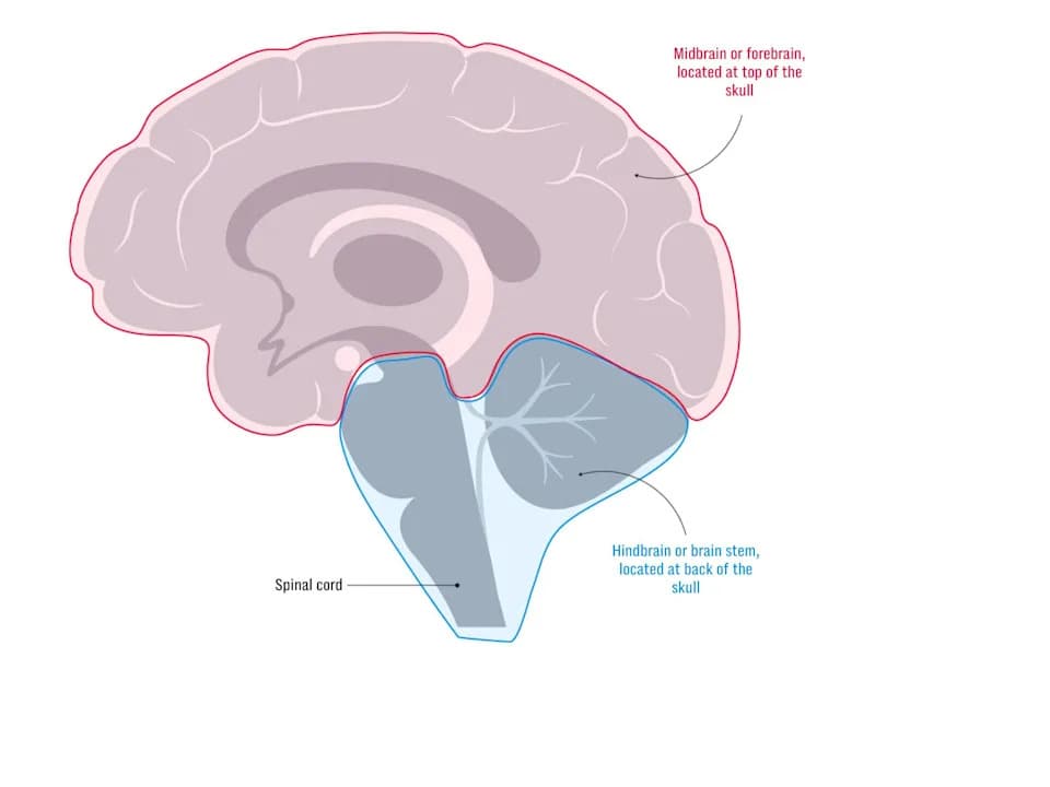
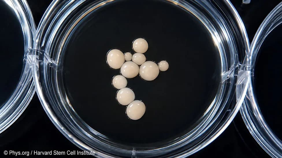
The team began by reprogramming donated human skin cells into neural tissue in the lab. To give that tissue room to grow, they engineered mice that develop without a cerebral cortex and hippocampus, creating a cavity with an estimated volume equivalent to about 14 million mouse cells. Newborn pups received several injections of roughly 100,000 human cells each.
Three months after transplantation the human grafts had become vascularized and filled much of the cavity with an estimated four million human neurons. Some of those neurons formed synaptic connections with mouse neurons and even with the spinal cord, demonstrating functional integration at a cellular level.
What the Grafts Looked Like And What They Did
Although the human tissue survived and integrated with host circuitry, it did not develop the layered structure of a mature human cortex. Instead, the grafts remained immature, resembling a human fetal brain at roughly mid-gestation. The mice did not show any measurable cognitive enhancement; externally they appeared broadly normal but walked cautiously and showed memory impairments that improved slightly after transplantation.
Experimental Findings And Applications
To demonstrate the model’s potential, researchers exposed some animals to prolonged oxygen deprivation (five hours at about 5% O₂) to mimic birth-related brain injury linked to cerebral palsy. Human neurons in the grafts were markedly vulnerable under these conditions, highlighting the model’s ability to reveal human-specific susceptibilities.
The team also identified von Economo neurons in the transplanted tissue — a specialized cell type previously observed only in postmortem human brains and implicated in frontotemporal dementia. That finding suggests the model could be useful for studying selective neuronal vulnerability in specific neurodegenerative diseases.
Ethics, Oversight, And Scientific Debate
Investigators report that the work proceeded under institutional review and followed relevant guidelines, including those of the International Society for Stem Cell Research (ISSCR); donors provided consent for transplantation. Nonetheless, the experiments reignite ethical questions about chimeric models and organoids — specifically, concerns about animal welfare, the possibility of emergent human-like activity in grafted tissue, and how to monitor and limit suffering.
There is also a scientific debate about model fidelity. Proponents note that in vivo models provide blood vessels and physiological signals that help human cells survive and mature; critics argue that growing human tissue in an engineered rodent cavity is artificial and may not faithfully reproduce human brain development. Some researchers advocate for prioritizing improved dish-based organoid models to reduce animal use and ethical friction.
Takeaway
This model offers a powerful, if ethically complex, platform to study human brain disorders in a living system — but important scientific and moral limits remain.
Sources: Nature, Stanford Medicine, Nuffield Council on Bioethics, The Guardian.
Help us improve.