Mouse experiments suggest peripheral immune activity—specifically dendritic cells in lymph nodes priming CD8 T cells—can drive Alzheimer’s‑like brain damage even when tau tangles remain. Removing peripheral dendritic cells eliminated excess brain T cells, reduced neurodegeneration and preserved cognition in mice. The findings point to new, potentially more accessible therapeutic targets outside the blood–brain barrier, but human studies are needed to confirm clinical relevance.
Peripheral Immune Cells May Drive Alzheimer’s‑Like Brain Damage, Mouse Study Finds

A stock image shows a doctor's hand in a surgical glove pointing at a brain scan image on a computer screen.
Scientists report new evidence that some immune activity linked to Alzheimer’s‑like brain damage may begin outside the brain, pointing to a potential new route for therapies that avoid the blood–brain barrier.
Study Overview
In a study published in Nature Neuroscience, researchers used mouse models that develop tau tangles—the twisted protein clumps that characterize Alzheimer’s disease and related tauopathies—to investigate how T cells become involved in neurodegeneration. The team focused on classical dendritic cells type 1 (cDC1), a dendritic cell subtype that can prime CD8 T cells, and asked whether these cells were acting inside the brain or in peripheral lymphoid tissue.
Key Findings
The investigators found very few cDC1 cells in brain tissue and no evidence that those present were directly engaging the T cells that accumulate after tau pathology appears. When dendritic cells were removed from peripheral locations including lymph nodes, the mice showed:
- Disappearance of the abnormally high numbers of brain T cells and a marked reduction in CD8 T cells;
- Substantial reduction in brain damage associated with neurodegeneration;
- Preservation of cognitive performance compared with untreated mice;
- No change in the amount of tau tangles present in the brain.
Together, these results suggest that dendritic cells in peripheral lymph nodes prime T cells that later migrate into the brain and contribute to neuronal injury—even when tau aggregates remain unchanged.
Proposed Mechanism and Implications
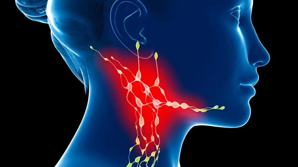
Although the researchers have not proven the full chain of events, they propose a plausible sequence: tau‑related neuronal damage releases brain-derived material that drains to cervical lymph nodes, where peripheral dendritic cells recognize antigenic material and prime CD8 T cells. Those T cells then traffic into the brain and exacerbate neurodegeneration.
The potential clinical significance is twofold. First, it identifies a peripheral immune pathway that may be easier to target than processes behind the blood–brain barrier. Second, it raises the possibility that therapies already used to modulate T cells in other diseases might be repurposed or adapted for neurodegenerative conditions—pending careful testing for safety and efficacy.
Caveats and Next Steps
Senior author Dr. David Holtzman (WashU Medicine) notes that human data already show increased T cells, including CD8 T cells, in brain regions with tau pathology and genetic links in the HLA locus that implicate T cell involvement. However, confirming a causal, therapeutically actionable role for peripheral dendritic‑cell priming in people will require trials that directly test whether manipulating T cells improves clinical outcomes.
The research team is now investigating whether blocking dendritic cell function later in life—around the time tau tangles begin to form—produces similar protective effects, and they are searching for the exact brain‑derived antigens or signals that prime peripheral dendritic cells.
Citation: Hao Hu et al., Priming of CD8+ T Cells by Peripheral Dendritic Cells Exacerbates Tau‑Mediated Neurodegeneration, Nature Neuroscience. DOI: 10.1038/s41593-026-02427-5.
Help us improve.