Researchers transplanted lab-grown human brain cells into mice engineered to lack a cortex and hippocampus, allowing the cells to integrate and mature inside the animals. The grafts connected to blood vessels and grew to nearly 4 million neurons, including rare von Economo neurons linked to frontotemporal dementia. Low-oxygen exposure caused walking difficulties, and scientists will test whether dementia-linked mutations increase vulnerability. The method could improve study of Alzheimer's, autism, epilepsy and schizophrenia while raising important ethical questions.
Lab-Grown Human Brain Cells Grafted Into Mice Provide New Model for Neurological Disease Research

Researchers have grafted lab-grown human brain cells into specially engineered mice to observe how the cells behave inside an intact, living brain. The study, published in Nature, reports that the transplanted human neurons integrated with the host tissue, connected to blood vessels and grew to nearly 4 million cells.
How the Study Was Done
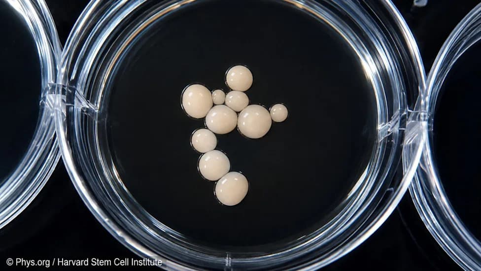
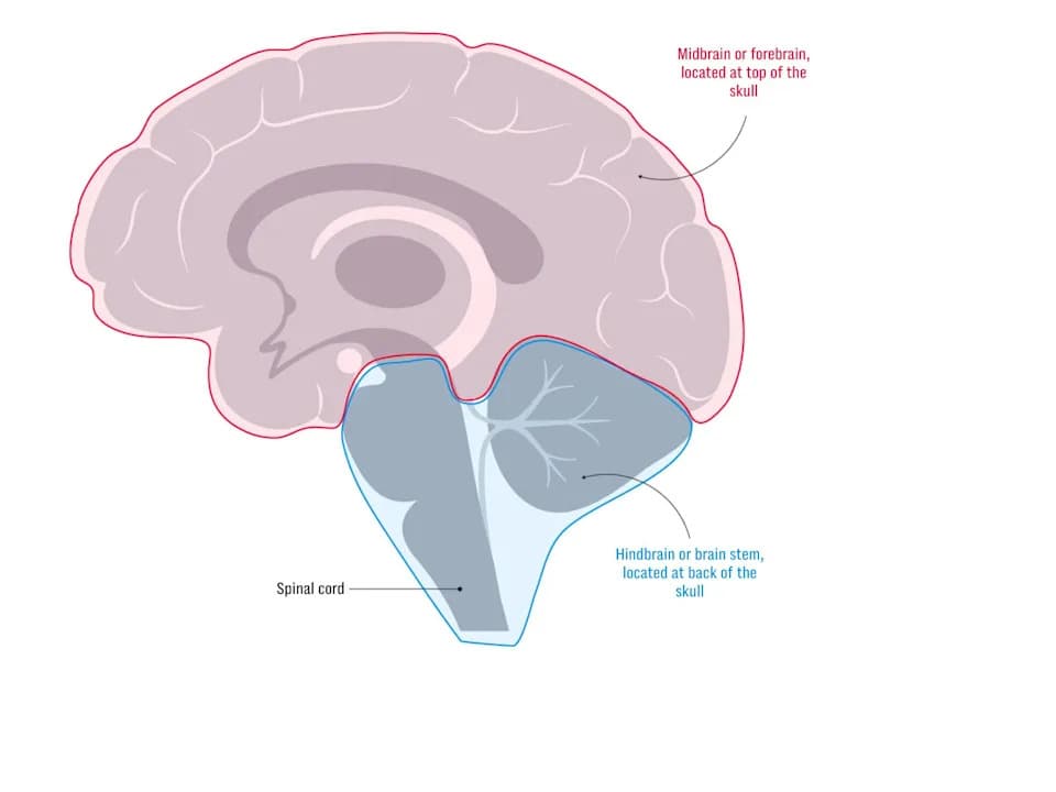
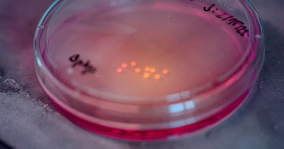
The research team bred mice that develop without a cortex or hippocampus, creating anatomical space for the human-derived tissue to expand. Human brain organoids placed into those sites thrived: they drew nutrients from the skull's fluids, established vascular connections, and matured over time into functioning human neurons within the rodent brain.
"For the past two decades there has been an effort to build models of the human brain outside the human body," said Dr. Sergiu Pașca, a professor at Stanford University. "This approach will not replace existing models, but it will give us access to aspects of human brain function that are otherwise very difficult to study."
Key Findings
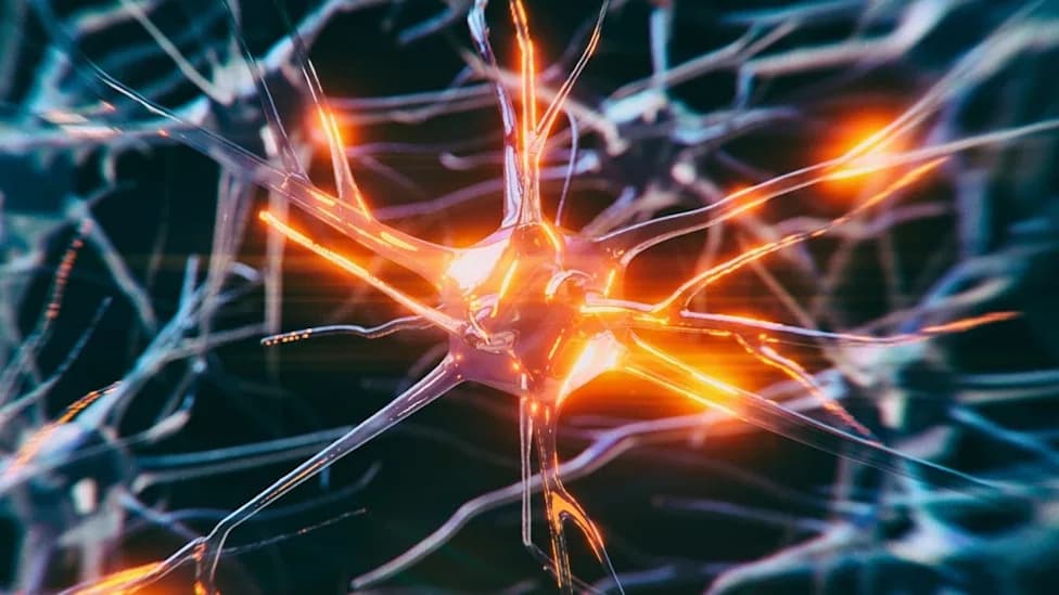
The grafted human tissue expanded to nearly 4 million neurons and included rare von Economo neurons, a cell type that is among the first to degenerate in frontotemporal dementia. To test vulnerability, some chimeric animals were exposed to prolonged low-oxygen conditions; those mice developed walking difficulties, suggesting the human-derived neurons were affected by the stressor.
Researchers plan to use this platform to test whether genetic mutations linked to frontotemporal dementia—or other neuropsychiatric and neurodegenerative disorders—make specific human neurons particularly susceptible to injury or disease.
Potential Applications and Considerations
Investigators say the method could advance study of Alzheimer’s disease, frontotemporal dementia, autism, epilepsy and schizophrenia by allowing scientists to observe living human neurons in a functioning brain environment. The approach may fill gaps left by cell cultures and organoids that lack full vascularization and long-term maturation.
Ethical and scientific caveats: The work raises ethical questions about chimeric models, and the authors emphasize careful oversight, strict limits on experimental design, and the need for reproducibility across labs. Translating findings from chimeric mice to human disease will require further validation.
Nature published the full study, which researchers say reflects years of breeding, refinement and collaboration to create a robust platform for studying human neurons in vivo.
Help us improve.