The University of Zurich-led studies found that transplanted human neural progenitor cells survived at least five weeks in stroke‑damaged mouse brains, differentiated into neurons, and formed connections with host circuitry, producing measurable motor improvements. Grafts also promoted angiogenesis, reduced inflammation, and helped restore blood‑brain barrier integrity. A related study found transplantation one week after stroke gave better initial graft survival than immediate transplantation. These are promising preclinical results, but safety, dosing, immune response, and delivery must be resolved before human trials.
Human Stem Cell Grafts Rebuild Neural Circuits and Improve Movement in Stroke-Damaged Mice
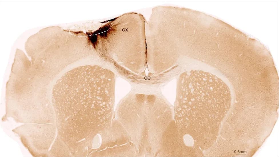
This image shows a coronal section through the mouse brain after stroke and neural stem cell transplantation. The dashed circle indicates the stroke area. The neurite projections of the transplanted human cells are stained in dark brown. Neurites extend locally into the cortex (CX) but also via the corpus callosum (CC) into the other brain hemisphere. (Image: UZH)
Researchers led by the University of Zurich report that human neural progenitor cells transplanted into brains damaged by experimentally induced ischemic stroke in mice survived for at least five weeks, matured into neurons, and integrated with host circuitry. The treated animals showed measurable motor improvements, and the grafts also appeared to promote blood-vessel growth, reduce inflammation, and help restore blood‑brain barrier integrity. These results come from two peer‑reviewed preclinical studies and do not yet demonstrate safety or effectiveness in humans.
How the Study Was Done
Teams at the University of Zurich, together with collaborators at the University of Southern California and Kyoto University’s Center for iPS Cell Research and Application, reprogrammed human somatic cells into induced pluripotent stem cells (iPSCs) and directed them toward a neural progenitor fate. Those neural progenitor cells were injected into the regions of mouse brains damaged by ischemic stroke.
The grafted human cells persisted throughout a five‑week analysis period reported in Nature Communications (September 2025). Most of the transplanted cells differentiated into neurons, and electrophysiological and anatomical data indicated these new neurons formed synaptic connections with existing mouse brain cells.
“We found that the stem cells survived for the full analysis period of five weeks and that most of them transformed into neurons, which actually even communicated with the already existing brain cells,” said Christian Tackenberg, Scientific Head of Division at the University of Zurich’s Institute for Regenerative Medicine.
Functional and Tissue Effects
Behavioral testing showed that mice receiving the grafts climbed more effectively and exhibited significantly improved gait compared with untreated controls. Movement recovery was assessed in part using AI‑assisted behavioral analysis. Beyond replacing lost neurons, the transplanted cells were associated with increased angiogenesis (blood‑vessel growth) in the injured cortex, signs of reduced inflammation, and improved blood‑brain barrier integrity.
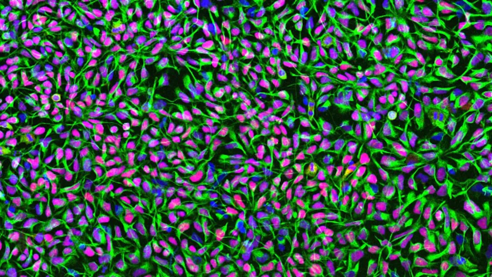
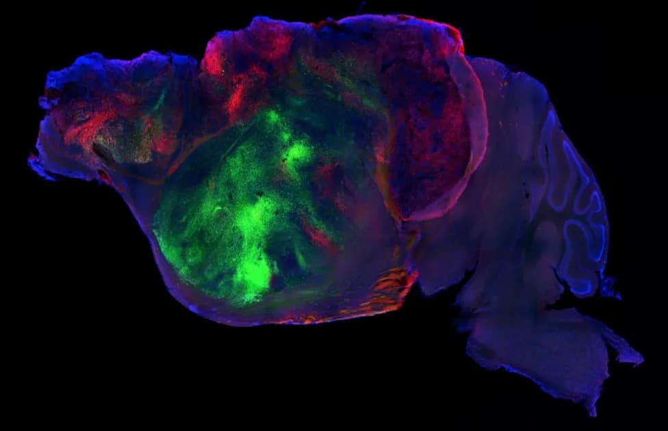
Human neural stem cells in culture. Cell nuclei are stained in blue, the neural stem cell-specific filament protein Nestin is shown in green, and the neural stem cell transcription factor Sox1 in red. (Image: UZH)
Timing, Delivery and Safety Considerations
A second study published online in Advanced Science (May 2025) reported that grafts transplanted one week after stroke had better initial survival than those placed immediately after injury, suggesting a potential therapeutic window that could allow time to prepare a tailored cell product after emergency care. Both studies used immunodeficient mouse strains to limit rejection of human cells, which facilitates study of graft behavior but does not reproduce the complexity of human immune responses.
The production protocol, developed with Kyoto University, avoided animal‑derived reagents—a choice that may ease some manufacturing and regulatory hurdles but does not establish clinical readiness. Direct intracerebral injection is invasive; the team is exploring less invasive endovascular delivery methods that would introduce cells via the bloodstream. Investigators also highlight the unresolved risk of uncontrolled cell growth and are developing a biological safety switch to eliminate transplanted cells if abnormal proliferation occurs.
What This Means and Next Steps
These studies provide encouraging preclinical evidence that human neural progenitor grafts can survive, mature, and functionally integrate in stroke‑damaged mouse brains while stimulating additional repair processes. However, mouse models cannot fully capture human stroke biology, immune responses, or long‑term safety. Before any clinical trials, researchers must demonstrate durable benefit, define safe dosing, refine delivery methods, confirm safety in more representative animal models, and address immune compatibility and manufacturing challenges.
Image caption: Human neural stem cells in culture. Nuclei stained blue; the neural stem cell filament protein Nestin shown in green; transcription factor Sox1 in red. (Image: UZH)
Help us improve.