Stanford researchers transplanted lab-grown human cortical organoids into newborn rats to observe human neuron maturation in vivo. After six months the grafts occupied about one-third of the implanted hemisphere, were vascularized by host blood vessels, and developed more complex branching than cells kept in culture. Sensory stimulation activated cells in the human graft, and optogenetic pairing with water rewards enabled rats to learn a light-linked behavior, demonstrating functional integration without obvious cognitive or motor changes.
Stanford Implanted Lab-Grown Human Brain Tissue Into Newborn Rats — The Grafts Wired Into Their Brains
Stanford Scientists Grew Human Brain Tissue in Mice—It Wired Into Their Nervous Systems
Stanford researchers transplanted lab-grown human cortical tissue into newborn rats to study how human neurons mature and function inside a living brain. In controlled experiments the implanted tissue expanded, received sensory input, and influenced learned behavior without producing obvious "human-like" animals.
How the Study Worked
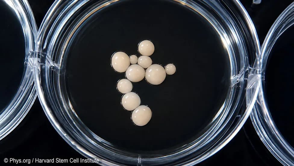
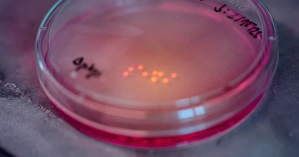
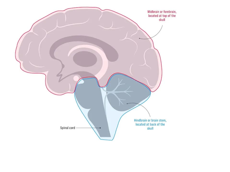
The team used cortical organoids — small, brain-like clusters grown from reprogrammed human cells — originally described in a 2022 Nature paper. Organoids model portions of the cerebral cortex, the brain's outer layer involved in memory, perception and thought. While useful in dishes, organoids lack blood flow, sustained immune support and natural sensory signals, which constrain maturation in vitro. Transplanting them into living brains provides a richer, more physiological environment.
Transplantation and Growth
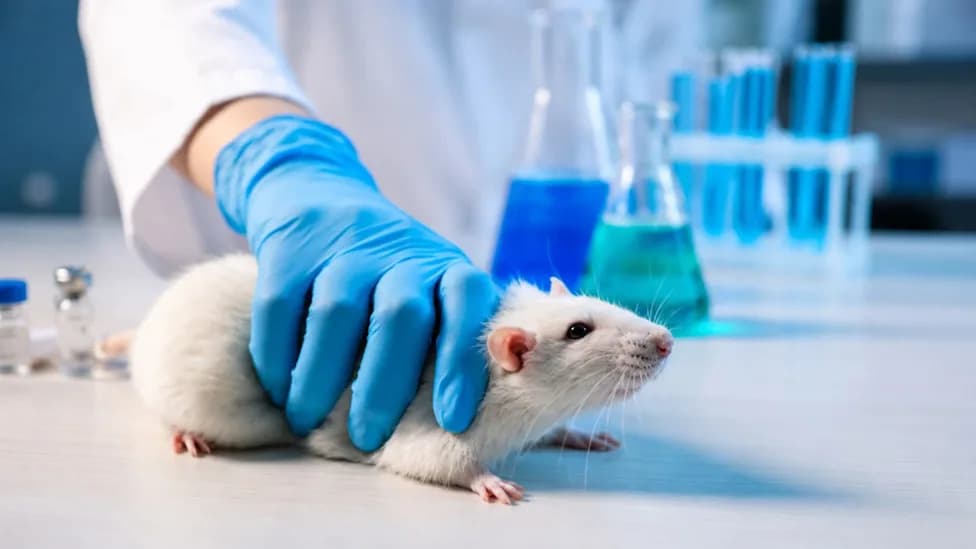
Researchers implanted organoids into the sensory cortex of rat pups two to three days after birth, when the animals' brains were still developing. Over six months the grafts expanded to occupy roughly one-third of the implanted hemisphere. Host blood vessels grew into the human tissue, supplying oxygen and nutrients, and the transplanted neurons enlarged and developed more elaborate branching than comparable cells left in culture.
Functional Integration
The human neurons became part of active neural circuits. When scientists puffed air across the rats' whiskers, cells inside the grafted tissue responded — evidence that sensory signals from the animal reached the transplant. Using optogenetics (activating targeted cells with light) and pairing light stimulation of the human neurons with water rewards, researchers trained the rats to approach a drinking spout when the light appeared. That learned behavior indicates the grafted human cells were able to influence host circuitry and behavior.
"This is the most advanced human brain circuitry ever built from human skin cells," said Sergiu Pașcă, the study's lead researcher. "Our platform provides, for the first time, behavioral readouts for human cells."
Implications and Cautions
The model offers a promising platform to study developmental and neuropsychiatric conditions such as autism, epilepsy and schizophrenia, and to test how human neurons respond to drugs or injury in an intact brain environment. The authors emphasize they did not create "human-like" rats: the animals showed no clear changes in memory, movement or seizure susceptibility. The work also raises ethical and regulatory questions about chimeric brain research that scientists and policymakers must address as the field advances.
Original publication: This story originally appeared in Men's Journal on Sep 19, 2026.
Help us improve.