Stanford researchers found that the human brain is formed from two evolutionarily distinct neural systems — a hindbrain (brain stem) and a front brain (midbrain and forebrain) — that developed independently and later joined spatially. Mouse gastrulation-stage studies revealed two non-overlapping progenitor lineages marked by Otx2 and Gbx2 with different chromatin states. Using these insights, the team derived functional human hindbrain motor neurons from pluripotent stem cells, enabling new disease models for SMA and ALS.
Stanford Finds Human Brain Is Two Ancient Nervous Systems Packaged Together
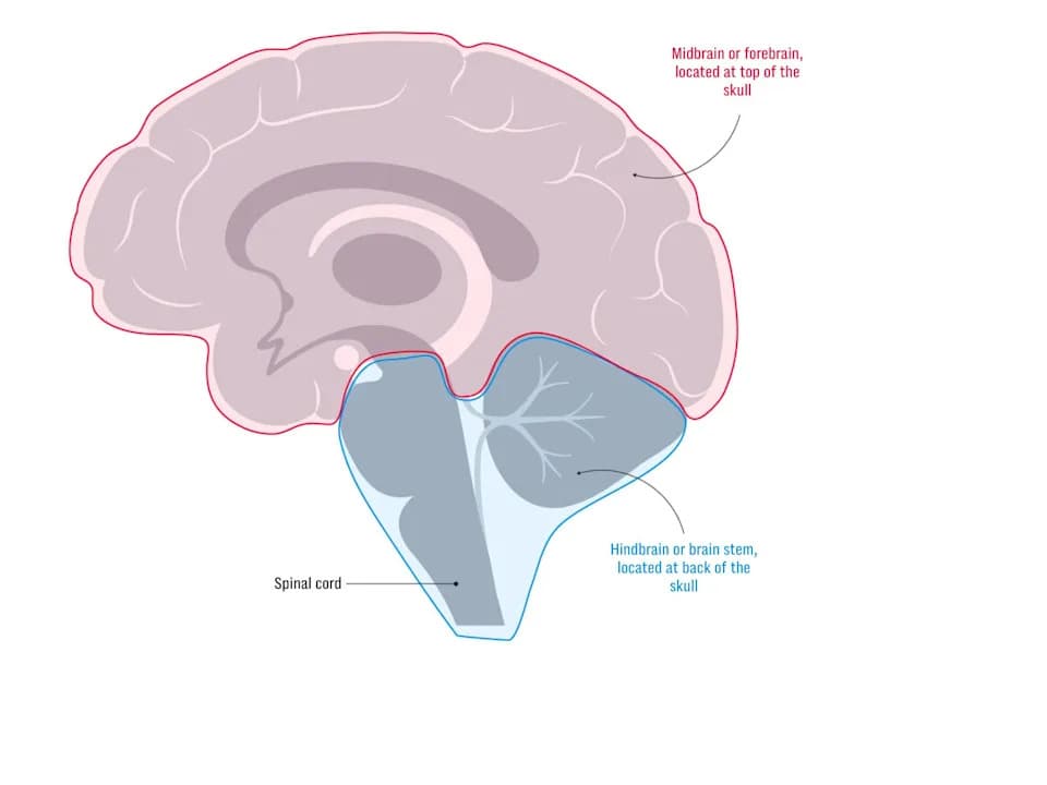
1909 Brain is two organs
Scientists at Stanford University report that the human brain is not a single homogeneous organ but the result of two ancient nervous systems that evolved separately and are now spatially joined and tightly integrated.
Separate Origins, One Functional Brain
Using mouse embryos at the gastrulation stage — when the body plan first emerges — researchers identified two mutually exclusive progenitor cell populations that give rise to different parts of the brain. One lineage, marked by the gene Otx2, becomes forebrain and midbrain structures responsible for higher cognition. The other, marked by Gbx2, forms the hindbrain (brain stem), which controls vital functions such as breathing, heartbeat, swallowing and sleep.
Detailed analysis showed the two progenitor populations have distinct chromatin (DNA packaging) states, effectively locking them onto separate developmental fates. Stanford researchers described this early bifurcation as like "travellers on parallel tracks that never cross," even though the mature brain integrates both systems seamlessly.
From Developmental Insight to Lab-Grown Neurons
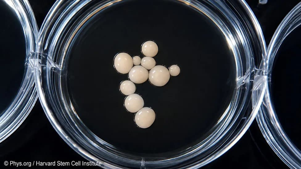
The team used this developmental insight to coax human pluripotent stem cells into functional hindbrain motor neurons in vitro, overcoming a longstanding barrier: hindbrain-derived neurons had been difficult to culture reliably compared with forebrain and midbrain cells. Producing large numbers of human brain-stem neurons in a petri dish creates new opportunities to model and study disorders that specifically affect the hindbrain.
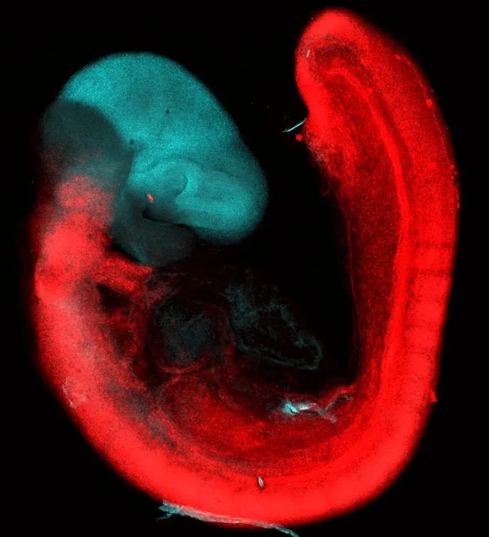
A brain scan of a 9.5-day-old mouse embryo shows the development of the two organs - Stanford Medicine/Loh Laboratory
Disease and Evolutionary Implications
Growing hindbrain motor neurons in vitro is expected to accelerate research into neurodegenerative diseases such as spinal muscular atrophy (SMA) and amyotrophic lateral sclerosis (ALS), where hindbrain motor neurons degenerate and impair swallowing, speech and breathing. Researchers are already using the new cell models to study how SMA kills hindbrain motor neurons and to screen potential drugs.
Comparative analysis suggests this two-part neural architecture is ancient: similar patterns were observed in acorn worms, distant relatives of vertebrates, implying separate neural systems may have existed around 500 million years ago and were later brought together by evolution.
"While the front and back of the brain are built from different sources, it is remarkable that they intimately connect with one another to form a functional brain," said Rayyan Jokhai, the study's first author. "Our ability to create large numbers of human hindbrain motor neurons in a petri dish offers a new approach to model these diseases."
The peer-reviewed findings appear in Nature Neuroscience and are led by Dr. Kyle Loh and colleagues at Stanford Medicine.
Why This Matters
This discovery reshapes our understanding of brain origin and organization, bridges developmental biology with disease modeling, and provides practical tools to study brain-stem disorders that previously lacked robust cellular models.
Help us improve.