The ISTA team generated a mouse cerebral cortex organoid from pluripotent embryonic stem cells and directly compared it with in vivo development using single-cell sequencing and MADM lineage tracing. The organoid produced the correct cell types and timely glial emergence but did not follow the strict, linear developmental sequence seen in living mice. Researchers conclude that missing stem cell niche elements—nearby cells, blood vessels, signaling molecules and mechanical cues—likely explain the difference and will be the focus of future improvements.
Lab-Grown Mouse Cerebral Cortex Matches Cell Types but Lacks In Vivo Timing
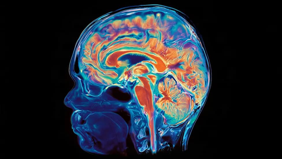
This Is the First Lab-Grown Mouse Cerebral CortexDragon Claws - Getty Images
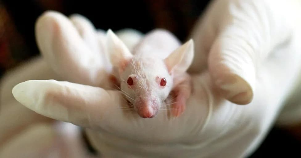
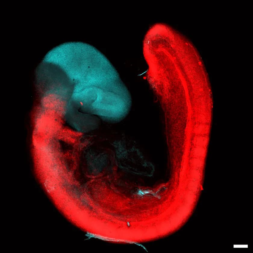
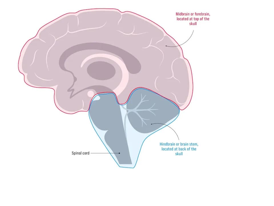
Researchers at the Institute of Science and Technology Austria (ISTA) have for the first time cultivated a structure resembling a mouse cerebral cortex from pluripotent embryonic stem cells, according to a new paper in Nature. The team built a cerebral cortex organoid to model normal brain development and to study disorders caused by developmental errors.
What the Team Did
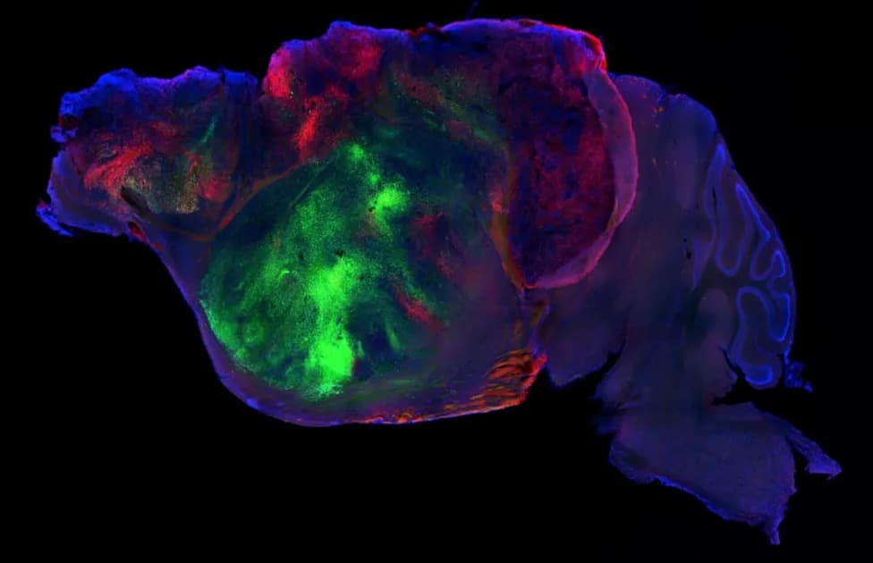
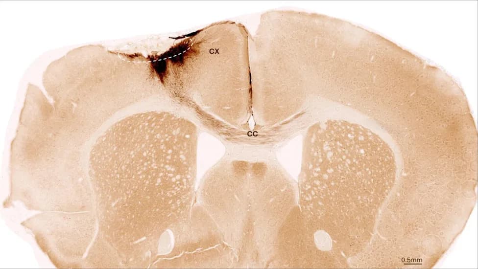
The researchers used mouse embryonic stem cells to generate a cortical organoid and then compared its development directly to that of living mice. They tracked cell types, proportions and timing with single-cell RNA sequencing and applied Mosaic Analysis with Double Markers (MADM), a genetic lineage-tracing technique that labels a stem cell and all of its descendants. Prior MADM work had defined a clear, linear developmental program for the mouse cortex in vivo; applying the same method to the organoid enabled a step-by-step comparison.
Key Findings
The organoid produced most of the expected cortical cell types, and glial cells appeared at roughly the correct stage. However, unlike the developing brain in a living mouse, the organoid did not follow the strict, linear sequence of proliferation, asymmetric neuronal production, then glial emergence. As ISTA senior author Simon Hippenmeyer observed, "The physical force of self-organization alone is apparently not enough." The authors concluded that important elements of the stem cell niche—nearby cells, blood vessels, signaling molecules, growth factors and mechanical cues—were absent or insufficient in the organoid.
Why This Matters
Understanding which aspects of brain development are faithfully reproduced in organoids and which are not is crucial for their use as experimental models. This work helps identify which current brain organoids can be used to study particular questions and highlights the need to recreate niche components to produce more faithful cortical models. Improved organoids could better model neurodevelopmental disorders such as microcephaly and macrocephaly and help test interventions.
Next Steps and Ethical Context
The authors suggest future work should probe and engineer components of the stem cell niche—for example, vascularization, signaling gradients and mechanical cues—to enforce a more ordered developmental program. As organoid technology advances, ethical discussions about complex brain-like tissues continue, but this study indicates that, at least for now, timing and microenvironment remain major hurdles to creating organoids that closely mirror in vivo brain development.
Bottom Line: The mouse cortical organoid reproduces many cell types but lacks the precise, linear developmental timing of an intact brain; recreating the stem cell niche is likely essential to close that gap.
Help us improve.