Key Points: A 36-mm sewing needle was found lodged in the left ventricle of a 35-year-old man who presented with painful urination and hematuria. CT and echocardiography revealed multiple metallic foreign bodies across the abdomen and pelvis; one needle had pierced the pericardium and entered the left ventricular cavity. Surgeons prioritised removal of the intracardiac needle due to risks of migration, rupture, and infection; the patient recovered. Clinicians suspect possible self-embedding behaviour and recommend multidisciplinary follow-up.
36-mm Sewing Needle Removed From Man’s Left Ventricle After He Sought Care For Blood In Urine
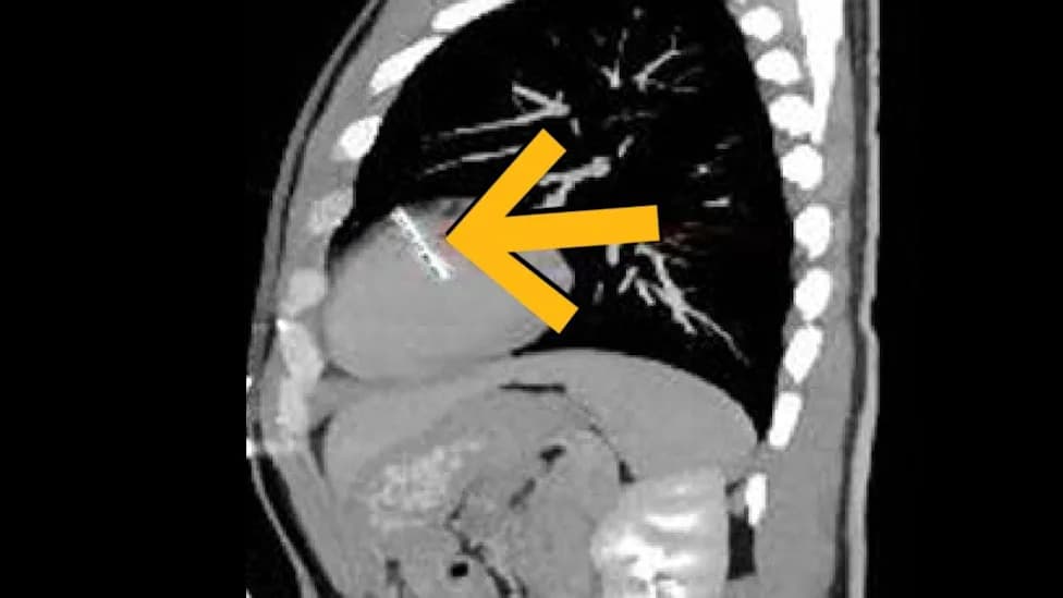
(Ren et al., BMC Cardiovascular Disorders, 2026)
Doctors in China have reported an extraordinary and potentially life-threatening case in which a 36-mm sewing needle was discovered lodged inside the left ventricle of a 35-year-old man who presented with painful urination and blood in his urine.
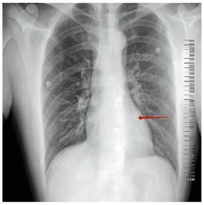
Chest X-ray demonstrating a linear metallic high-density shadow projected over the precordial region (red arrow). (Ren et al.,BMC Cardiovascular Disorders, 2026)
The patient, described as having an intellectual disability, arrived with normal vital signs and no obvious cardiac complaints. Routine imaging, however, revealed a linear metallic shadow over the precordial region. Further computed tomography (CT) of the chest, abdomen and pelvis uncovered multiple metallic foreign bodies distributed throughout his body — in the abdominal wall, pelvis, bladder wall and lumen, hip bone and muscles, and the penis — including one needle that had penetrated the pericardial sac and extended into the left ventricular cavity.
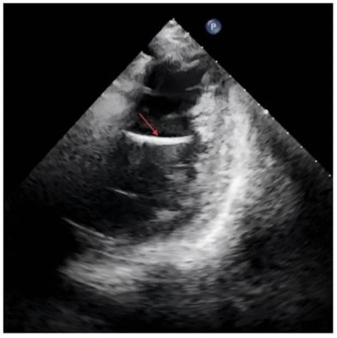
Transthoracic echocardiography confirming a 36-mm linear hyperechoic structure (red arrow) within the left ventricular cavity, which moved synchronously with the ventricular wall. (Ren et al.,BMC Cardiovascular Disorders, 2026)
Imaging and findings
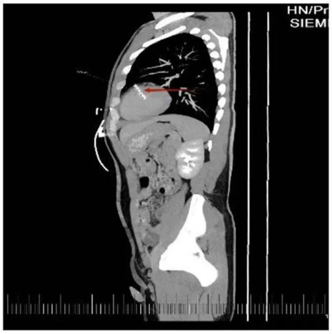
CT scan of the needle in the heart. (Ren et al.,BMC Cardiovascular Disorders, 2026)
Chest X-ray showed a linear, high-density metallic shadow projected over the precordium. Transthoracic echocardiography confirmed a 36-mm linear hyperechoic structure moving synchronously with the ventricular wall. CT slices identified multiple linear metallic foreign bodies across the pelvic and abdominal cavities (Ren et al., BMC Cardiovascular Disorders, 2026).
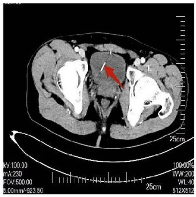
CT images identifying multiple linear metallic foreign bodies distributed within the pelvic cavity. (Ren et al.,BMC Cardiovascular Disorders, 2026)
Clinical course and surgery
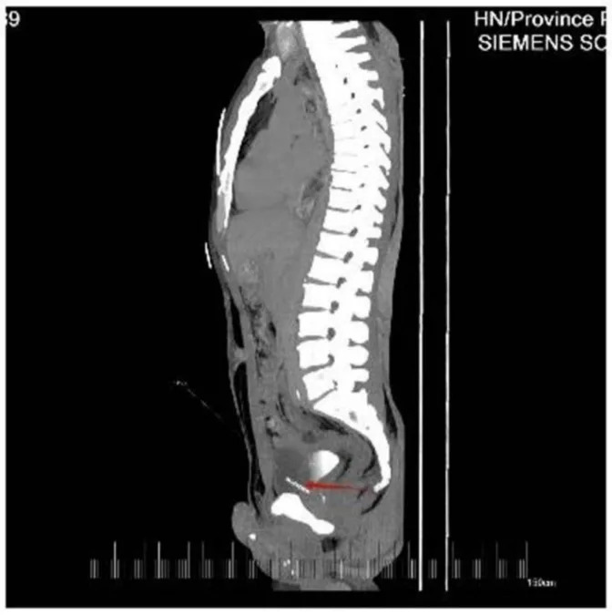
Other systemic foreign bodies in the abdominal-pelvic region. (Ren et al.,BMC Cardiovascular Disorders, 2026)
Because of the risk that the intracardiac needle could migrate, cause cardiac rupture, or lead to infection, the surgical team prioritised removal of the needle from the left ventricle. Preoperative assessment noted mild left ventricular dilation and early structural abnormality. Intraoperatively, clinicians also observed needles penetrating the bladder wall with stone-like encrustations; these bladder foreign bodies were deemed high risk for hemorrhage and urinary leakage and were scheduled for staged removal after the cardiac procedure. The intracardiac needle was removed successfully, and the patient recovered well.

Subscribe to ScienceAlert's free fact-checked newsletter
Origin and psychiatric considerations
Neither the patient nor his family could explain how the needles entered his body. Treating clinicians at Hunan Provincial People's Hospital noted that self-embedding — the deliberate insertion of objects into the body — is a possible explanation. The patient's record suggested a prior episode two years earlier in which two needle-like objects were removed from his abdomen, but recent treating teams lacked access to those earlier records, and whether prior incidents were accidental or deliberate remains unclear.
Context and implications
The authors highlight that intracardiac foreign bodies, particularly those resulting from presumed self-insertion, can be clinically occult. A 2024 review identified only 34 reported cases since 1967 of needles introduced into the heart as a form of self-injury. Needles in the heart may remain asymptomatic for months or years, making high clinical suspicion and careful imaging essential when patients are at risk or present with unexplained foreign bodies.
"Presumed self-insertion of cardiac foreign bodies in patients with cognitive impairment can be clinically occult, requiring a high index of clinical suspicion when a definitive history is difficult to obtain," the treating team concluded (Ren et al., BMC Cardiovascular Disorders).
The case report — published with written informed consent from the patient's father — underscores the need for multidisciplinary care, including surgical, urologic and psychiatric assessment and follow-up for patients found with multiple systemic foreign objects. Images accompanying the report demonstrate the linear metallic shadow on chest X-ray, CT images of pelvic foreign bodies, and echocardiography showing the hyperechoic intracardiac structure.
Help us improve.