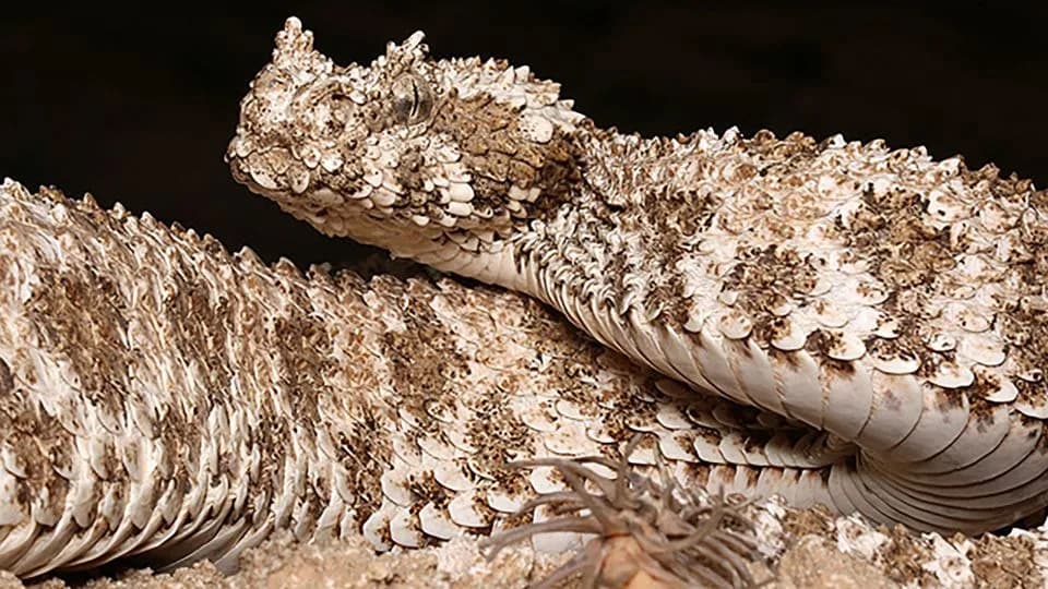
The spider-tailed horned viper (Pseudocerastes urarachnoides) uses a tail tip that mimics a spider to lure prey. μCT scans of the 1968 Field Museum holotype show no special bones at the tail tip—the illusion is produced entirely by modified, keratinized scales. Researchers call this an extreme example of skin evolution and note it highlights how soft-tissue adaptations can be invisible in the fossil record. The study appears in The Anatomical Record (Georgalis et al., 2026).
Spider-Tailed Viper’s “Spider” Is All Skin: μCT Scans Reveal No Bone Underlying the Illusion
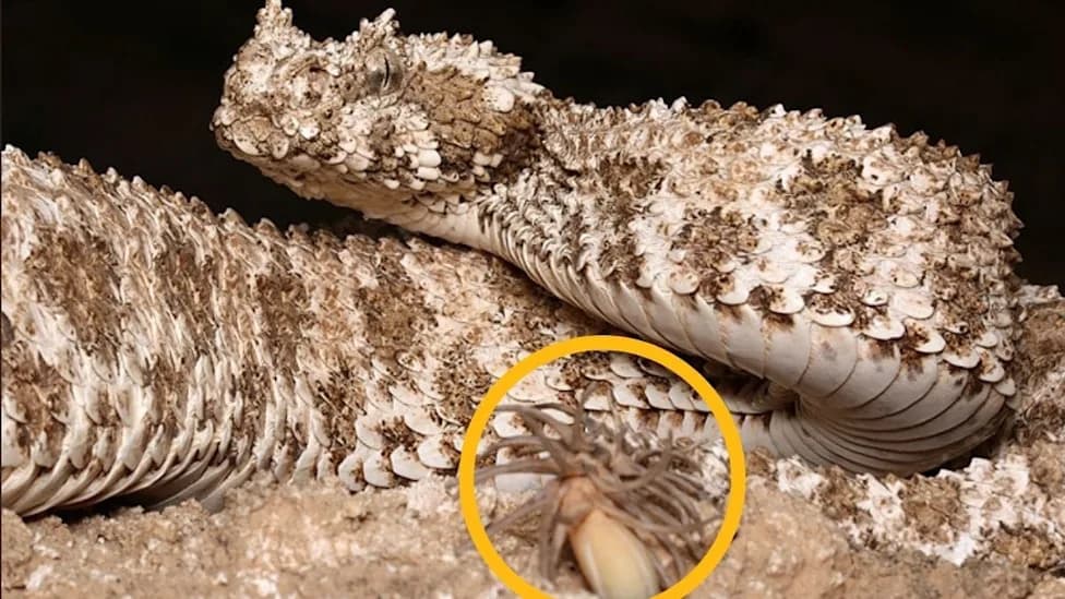
(Omid Mozaffari/Wikimedia Commons, Public Domain)
The spider-tailed horned viper (Pseudocerastes urarachnoides) of Iran is famous for a tail tip that convincingly mimics a spider. A new study using high-resolution micro-computed tomography (μCT) of the species' 1968 Field Museum holotype reveals the lifelike lure is formed entirely from modified skin and scales, not from any special bony anatomy.
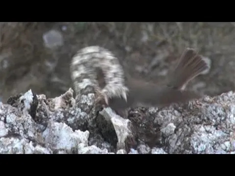
YouTube Thumbnail
Background. When the unusual 1968 specimen first drew scientific attention, researchers debated whether the tail growth was a deformity, tumor or parasite. After additional specimens were found and the species was formally recognized in 2006, the spider-like tail was established as a consistent trait rather than a one-off anomaly.
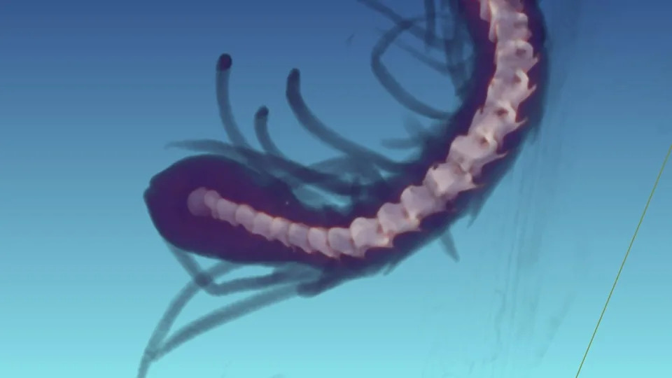
A micro-CT scan showing the normal bones inside the snake's bizarre tail. (Image courtesy of Sara Ruane and Stephanie Smith)
What the μCT scans show. Led by Georgios Georgalis (Polish Academy of Sciences) and colleagues, researchers scanned the holotype at the Field Museum's XCT Lab to examine the tail vertebrae and surrounding tissues. The scans showed normal bony anatomy at the tail tip: there are no special bones or skeletal modifications that form the spider-like filaments. Instead, the entire structure is produced by keratinized skin—elongated and fused scales that create radiating, flexible filaments.
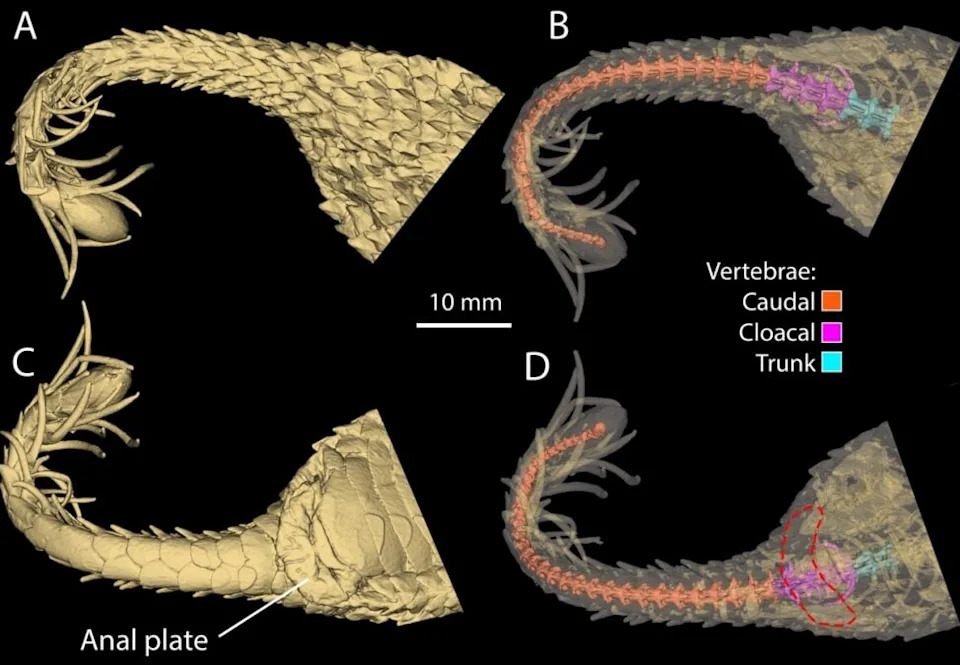
3D visualization of the tail vertebrae and surrounding soft tissues. (Georgalis et al.,The Anatomical Record, 2026)
“We expected there might be some obvious bony element in the tail or something at the end that alluded to the external features, but nope! You only get the spider tail from the soft tissue with no underlying bony structure, it is all keratin, just like our fingernails.” — Sara Ruane, Field Museum
Function and behavior. The viper uses this tail tip in a hunting strategy called caudal luring. Concealed against rocky terrain and nearly motionless, the snake intermittently twitches its tail so the filaments move like a small spider. Insectivorous birds and other prey that approach the apparent spider can become the snake's next meal. While caudal luring occurs in many snakes, the spider-tailed viper’s realistic multi-filament lure is unique in its effectiveness.
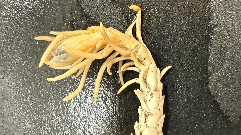
A close-up of the tail tip. (Georgalis et al.,The Anatomical Record, 2026)
Evolutionary and paleontological implications. Because the adaptation is made of soft tissue, it would rarely be preserved in fossils. A fossil relative of this species would likely show no trace of the elaborate lure, demonstrating how much soft-tissue variation may be invisible in the fossil record. The researchers describe this as an extreme example of integumentary evolution—a dramatic modification of scales rather than bones.

Subscribe to ScienceAlert's free fact-checked newsletter
Study details. The findings and three-dimensional visualizations of the tail vertebrae and soft tissues are reported in The Anatomical Record (Georgalis et al., 2026). The holotype images and scans were produced with assistance from the Field Museum's XCT Lab.
Note: Behavioral footage and prior studies helped establish the function and prevalence of this adaptation; the μCT work provides the first comprehensive internal anatomical assessment of the holotype.
Help us improve.