The study led by Professor Minah Suh shows that blocking PD-L1 in the brains of Alzheimer’s-model mice restored microglial responsiveness and reduced neuronal hyperactivity. Researchers observed elevated PD-1 on microglia and increased PD-L1 on astrocytes in the model. Direct intracerebral delivery of a PD-L1 antibody produced stronger benefits than systemic dosing. The findings are preclinical and highlight both the potential of immune-targeting strategies and the significant challenges of safely delivering antibodies to the human brain.
Blocking PD-L1 in Mouse Brains Restores Microglial Function and Lowers Neuronal Hyperactivity in Alzheimer’s Model
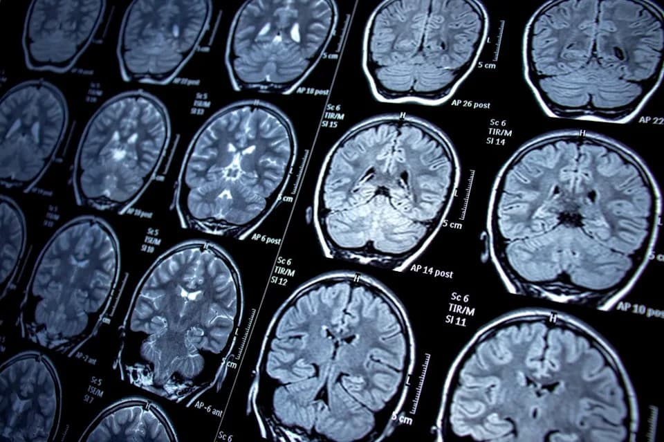
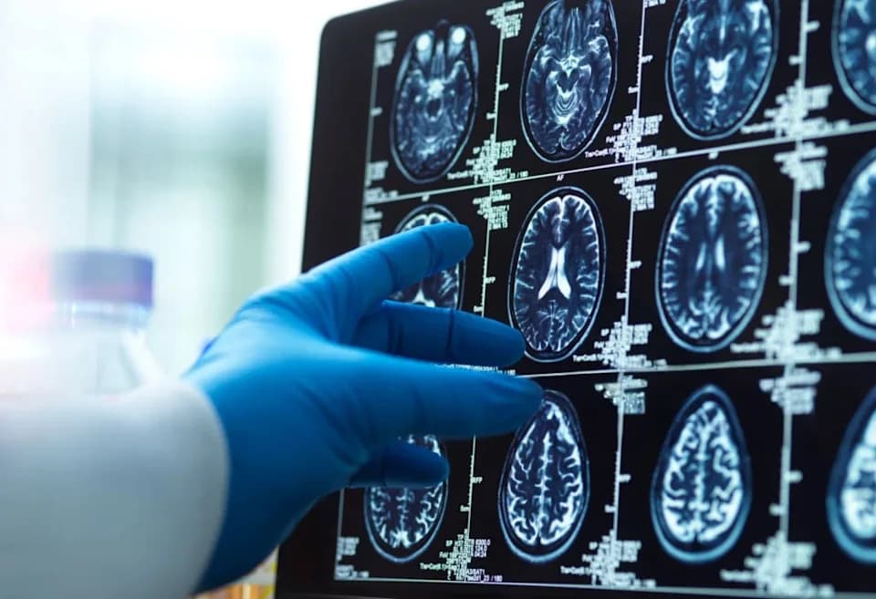
Stock image: MRI scans of a human brain are seen on screens.
Researchers report a promising preclinical approach that partially restores the brain's innate immune response and reduces abnormal neuronal hyperactivity linked to Alzheimer’s disease.
The study was led by Professor Minah Suh of the Department of Biomedical Engineering at Sungkyunkwan University, in collaboration with biotechnology company IMNEWRUN and Professor Ho-Keun Kwon's team at Yonsei University College of Medicine. The team focused on microglia, the brain's resident immune cells that detect damage and help preserve a healthy neural environment.
In healthy brains, microglia act as first responders to injury and help maintain local homeostasis. In Alzheimer’s disease, however, microglial responses become impaired and neurons can show excessive, damaging activity. The new study, published in Science Advances, links this dysfunction to altered signaling of the immune checkpoint proteins PD-1 and PD-L1.
Using an Alzheimer’s-like mouse model, the researchers found increased PD-1 expression on microglia and elevated PD-L1 on astrocytes, the supportive cells that contribute to the brain's chemical environment. To test the effect of this signaling imbalance, investigators administered a PD-L1–blocking antibody directly into the brains of the model mice and used advanced in vivo microscopy to monitor live brain cells.
The intervention produced notable results: microglia regained more normal surveillance and response behaviors around damaged tissue, and neuronal hyperactivity—previously elevated in the model—was reduced. Effects were stronger when PD-L1 was targeted within the brain than when the antibody was delivered systemically.
“This study indicates that immune cells (specifically microglia) may be directly involved in the process and is likely to increase interest in immune-mediated research,” said Dr. John Showalter, chief operating officer of Linus Health, who was not involved in the study. He also pointed to epidemiological evidence linking reduced dementia risk with certain vaccinations as one of several clues implicating immune mechanisms.
While the findings add to growing interest in immune-modifying strategies for Alzheimer’s, the authors and independent experts emphasize important caveats. The work was performed in mice engineered to model aspects of Alzheimer’s pathology, so it does not demonstrate that PD-L1 blockade will treat or cure human Alzheimer’s disease. A major translational challenge is safe and effective delivery of antibody therapies to the human brain; direct injection into cerebrospinal fluid carries significant risks and alternative delivery methods must be developed and tested.
The paper is cited as: Taeyoung Park et al., Targeting Glial PD-1/PD-L1 Restores Microglial Homeostasis and Reduces Neuronal Hyperactivity in an Alzheimer’s Disease Model, Science Advances (2026). DOI: 10.1126/sciadv.adx0731.
Takeaway: The study provides strong preclinical evidence that adjusting PD-1/PD-L1 signaling in the brain can restore microglial function and dampen pathological neuronal activity, highlighting immune pathways as a promising area for future Alzheimer’s research—while underscoring the hurdles to translate these findings to human treatments.
Help us improve.