UCL researchers trained an AI model on 11,089 MRI scans from over 8,500 patients in four countries to predict which brain tumours would enhance on contrast MRI without administering gadolinium. On a validation set of 1,100+ non-contrast images the model achieved 83% overall accuracy, with 92% sensitivity for enhancing tumours and 74% specificity for non-enhancing tumours. It performed best for meningioma but was less reliable in children; researchers see immediate use as a triage or decision-support tool rather than a full replacement for contrast-enhanced MRI.
AI May Spare Some Brain Tumour Patients From MRI Contrast Dye — Study
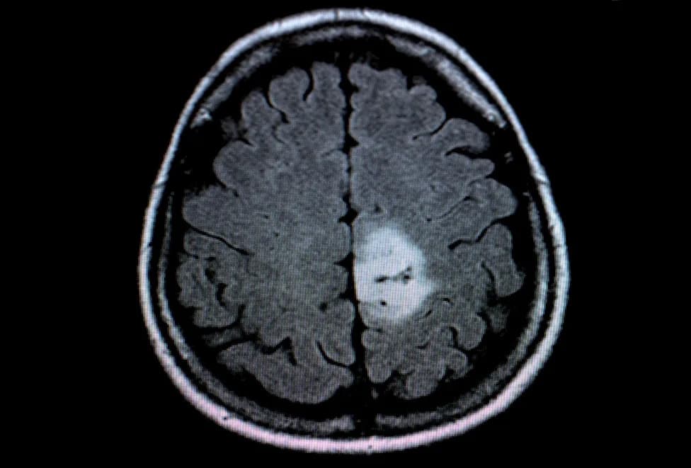
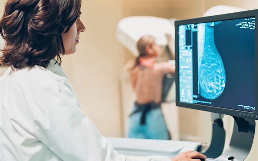
Brain tumour patients could be spared contrast dye injections during MRI scans with the help of artificial intelligence (AI). Rolf Vennenbernd/dpa
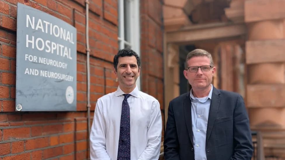
Researchers at University College London (UCL) have developed an artificial intelligence (AI) tool that could help identify which brain tumours would show enhancement on MRI scans without injecting gadolinium-based contrast dye. The approach aims to reduce unnecessary injections, avoid extra hospital visits and limit potential environmental contamination from gadolinium.
Why This Matters
Gadolinium is commonly injected during MRI to improve image clarity and help diagnose brain lesions. While widely used, the long-term effects of gadolinium exposure on the body remain uncertain, and trace amounts have been detected in sewage, surface and drinking water — sometimes far from MRI facilities. These concerns have motivated research into alternatives that could reduce reliance on contrast agents.
What The Study Did
UCL researchers trained an AI model using 11,089 MRI scans from more than 8,500 patients across the UK, US, the Netherlands and Nigeria. The model was designed to predict, from non-contrast MRI images alone, which regions of the brain would have enhanced signal if gadolinium had been given.
Key Findings
On a validation set of more than 1,100 non-contrast images, the algorithm correctly predicted whether a tumour would brighten on contrast-enhanced scans 83% of the time overall. The model detected 92% of tumours that did enhance (sensitivity) and correctly identified 74% of tumours that did not enhance (specificity). Performance was highest for meningioma — a common, typically benign brain tumour — and was lower in paediatric cases.
Clinical Implications and Limitations
The researchers emphasize that the model is not yet sufficient to replace contrast-enhanced MRI, particularly for children where accuracy was reduced. However, it could be valuable as a triage or decision-support tool to flag lesions likely to enhance, helping clinicians prioritise who needs contrast. Such use could reduce the number of patients needing a second visit for contrast-enhanced imaging and speed up care for some patients.
“AI innovation that could reduce the need for contrast dye injections during MRI scans for some patients is welcome,” said Dr Karen Noble, Director of Research and Policy at Brain Tumour Research. “We look forward to understanding how this model could be used in future to improve care, minimise side effects and help inform treatment decisions for brain tumour patients.”
Next Steps
Further validation in larger and more diverse paediatric cohorts, additional tumour types and real-world clinical workflows will be necessary before the tool can be used routinely. If corroborated, the approach could reduce exposure to gadolinium for many patients and mitigate environmental release from medical imaging.
Help us improve.