Researchers developed Pathology-CoT, a method that trains vision-language models using pathologists’ navigation patterns and short rationales, and built a prototype called Pathology-o3. Trained on logs from eight pathologists, the system scans whole-slide images at low resolution, selects regions of interest and analyzes high-resolution crops. In tests on lymph node slides from colorectal cancer cases, Pathology-o3 showed very high sensitivity (100% initial; 97.6% on an external set) but produced more false positives than some baseline models. The approach improved multiple VLMs, and follow-up trials will test whether pathologists working with the tool are faster or more accurate.
AI That Searches Like a Pathologist Boosts Cancer Detection — With Caveats
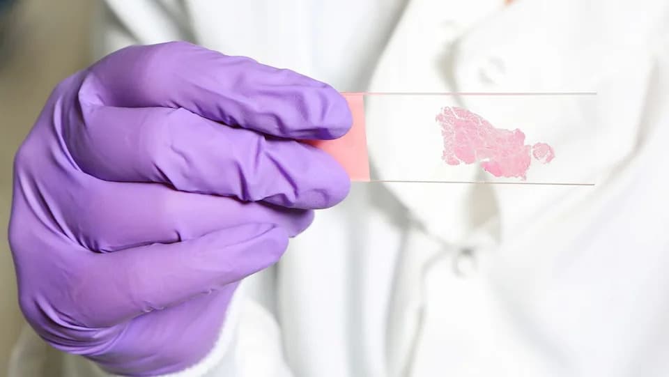
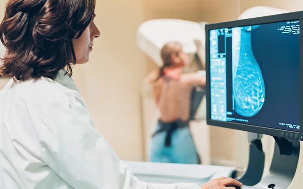
In a new study, researchers trained AI to screen tissue-sample slides similar to how a human pathologist would. | Credit: Nicola Tree via Getty Images
Researchers have taught AI to examine whole-slide pathology images the way human pathologists do — scanning broadly, zooming in on suspicious areas and pausing over regions that merit closer inspection. A team led by Zhi Huang (University of Pennsylvania) describes a training method, Pathology-CoT (chain of thought), that converts pathologists’ navigation behavior and short rationales into supervision for vision-language models (VLMs). The group used this approach to build a prototype called Pathology-o3.
The motivation is simple: whole-slide images can contain billions of pixels while the signs of cancer may occupy only tiny, hard-to-find regions. Many AI systems work on preselected patches or uniform tiles; by contrast, a human expert scans at low resolution, then zooms and lingers on areas that look suspicious. Huang compared the process to a search-and-rescue helicopter that first surveys the landscape and then swoops in.
How the System Was Trained
The researchers logged slide navigation and magnification changes from eight pathologists using a custom tool. The raw logs were noisy — with incidental drifts, overshoots and small adjustments — so the team filtered the data to emphasize deliberate attention (sustained pans and lingering views) and validated those traces against eye-tracking records.
For each inspected region, a VLM generated a short rationale describing why the view warranted attention and what features were visible. Pathologists reviewed these rationales, accepting, editing or rejecting them; that feedback became additional labeled training data. Using this combined navigation-and-rationale training, the team trained models to prioritize regions for high-resolution review.
Prototype Performance and Comparison
Pathology-o3 first scans a slide at low resolution, selects candidate regions using the navigation-trained model, then passes high-resolution crops of those regions to a VLM for analysis. The researchers compared Pathology-o3 with other generalist models (including OpenAI’s o3) on lymph node slides from colorectal cancer cases (some with metastatic deposits annotated by pathologists).
Pathology-CoT trains algorithms to scan over a whole slide and then return to regions of interest for a closer look. | Credit: Universal Images Group via Getty Images
Key results from the reported tests:
- On the initial test set, Pathology-o3 achieved 100% sensitivity (it detected all slides containing cancer); among slides it flagged as positive, 15.5% were actually negative (false-positive rate among positives).
- By comparison, OpenAI o3 achieved 87.5% sensitivity and had 53.3% of its positive calls turn out to be negative.
- On an independent external dataset, Pathology-o3 retained high sensitivity (97.6%) but showed a higher false-positive fraction among flagged slides (37.1%), illustrating performance shifts across data sources.
The authors intentionally tuned the prototype to favor overcalling (more false positives) rather than missing cancers. As a result, the system is better suited as a prescreening aid that draws a pathologist’s attention to specific regions rather than as an autonomous diagnostic tool.
Limitations and Next Steps
Experts not involved in the study cautioned that the system is not yet precise enough to diagnose patients independently. The study did not measure whether pathologists assisted by Pathology-o3 become faster or more accurate when reading slides; the team has planned follow-up experiments to test pathologists on identical cases with and without the tool, measuring detection rates and time-to-diagnosis.
Other limitations include reliance on single-slide reads (clinical diagnosis typically integrates multiple slides, special stains and patient history) and the modest number of contributing pathologists (eight) used to generate navigation traces. The researchers also applied Pathology-CoT to several existing VLMs and observed consistent performance improvements, suggesting navigation-based supervision can benefit multiple model architectures.
Practical Outlook: Pathology-CoT and Pathology-o3 show promise as prescreening or triage tools that flag candidate regions for human review. Larger, multicenter trials are needed to evaluate accuracy, speed, workload impact and the operational costs of false alarms before clinical deployment.
This article is for informational purposes and is not medical advice.
Help us improve.