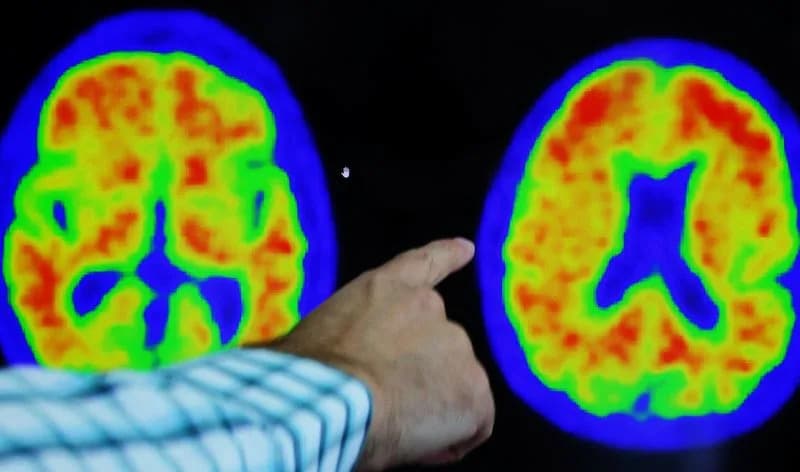
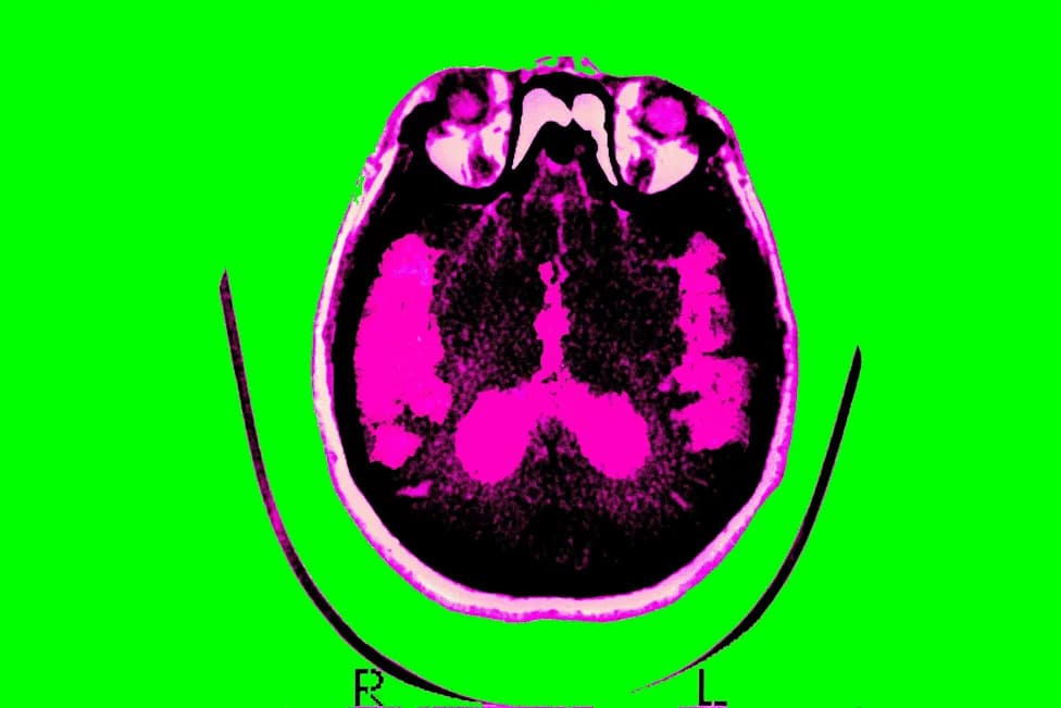
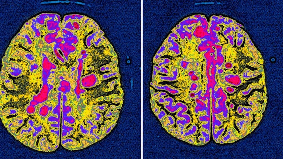
Researchers in the Netherlands report that lipid-filled 'foamy' microglia are associated with more severe progression of multiple sclerosis. Analysis of post-mortem brain tissue from 28 secondary progressive MS patients and 10 controls showed distinct molecular signatures around lesions where these cells accumulate. Blocking an enzyme active in foamy microglia improved tissue repair in a mouse model, and related lipids were detected in cerebrospinal fluid, pointing to potential therapeutic targets and biomarkers. Clinical trials and longitudinal studies are needed to confirm these findings.
Foamy Microglia May Drive Severe Multiple Sclerosis Progression — Potential Treatment Target and Biomarker Identified
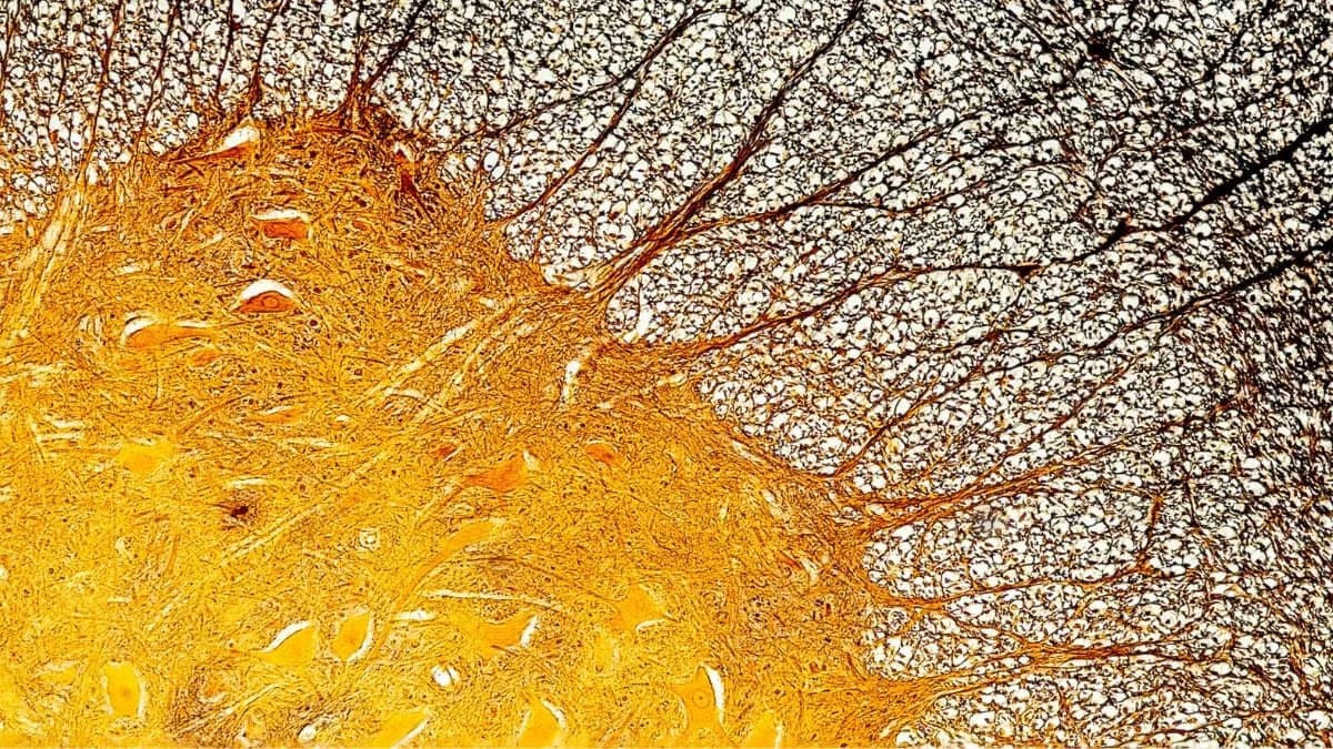
A cross-section of the spinal cord showing neurons and glial cells. (Juan Carlos Juarez Jaramillo/iStock/Getty Images Plus)
A diagnosis of multiple sclerosis (MS) often brings uncertainty about how the disease will progress. New research from the Netherlands points to a potential mechanism behind the most aggressive forms of MS — and suggests a possible therapeutic target and biomarker.
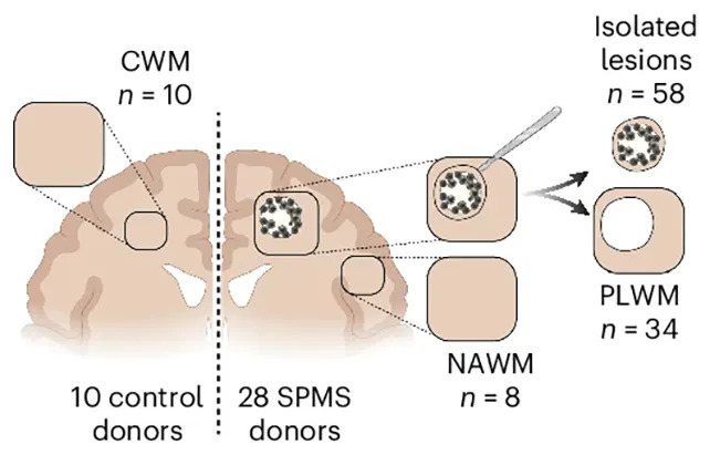
The researchers compared brain tissue samples collected from people with and without severe MS, looking at the damage (lesions) on nerve cells. (van der Vliet et al.,Nat. Neurosci., 2026)
Key Findings
MS damages nerve cells by stripping away myelin, the fatty insulating layer that ensures reliable nerve signaling. The study reports that in the most severe cases, microglia — the brain's resident scavenger immune cells — become overloaded with lipid droplets and take on a 'foamy' appearance. These lipid-laden microglia appear dysfunctional and may drive worse tissue damage and inflammation rather than promoting repair.
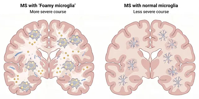
Severe MS was associated with fat-laden, 'foamy' microglia. (Netherlands Institute for Neuroscience)
What The Researchers Did
The team analyzed post-mortem brain tissue from 28 people with secondary progressive MS and compared these samples to tissue from 10 donors without MS. Using complementary molecular profiling methods, they mapped proteins, lipids and active genes in areas surrounding MS lesions. The study found a correlation between higher numbers of foamy microglia and more severe disease progression, along with a distinct molecular signature of proteins and enzymes around those lesions.

Subscribe to ScienceAlert's free fact-checked newsletter
"We found that patients with large numbers of these foamy microglia had a more severe disease course more frequently," said molecular physiologist Daan van der Vliet of Leiden University. "It does not appear to be simply about the inflammatory response alone."
Experimental Evidence From Mice
To test causality, the researchers used a mouse model of MS and blocked an enzyme highly active in foamy microglia. Mice treated this way showed improved tissue healing, strengthening the link between lipid-overloaded microglia and worse MS outcomes.
Implications And Next Steps
The authors detected lipids associated with foamy microglia in cerebrospinal fluid (CSF), suggesting these molecules could become measurable biomarkers to identify patients at higher risk of rapid decline. However, the team cautions that the research is still early: clinical trials in people with MS are required to confirm whether targeting microglial lipid processing can alter disease progression. Additional longitudinal studies will also be necessary to understand how unrepaired lesions evolve over time.
"These cells are probably trying to do something good: clearing up damage," van der Vliet added. "But they become overloaded, so to speak. As a result, they can no longer effectively contribute to repair."
The study is published in Nature Neuroscience. While promising, translation into human treatments and diagnostic tests will require further validation.
Help us improve.