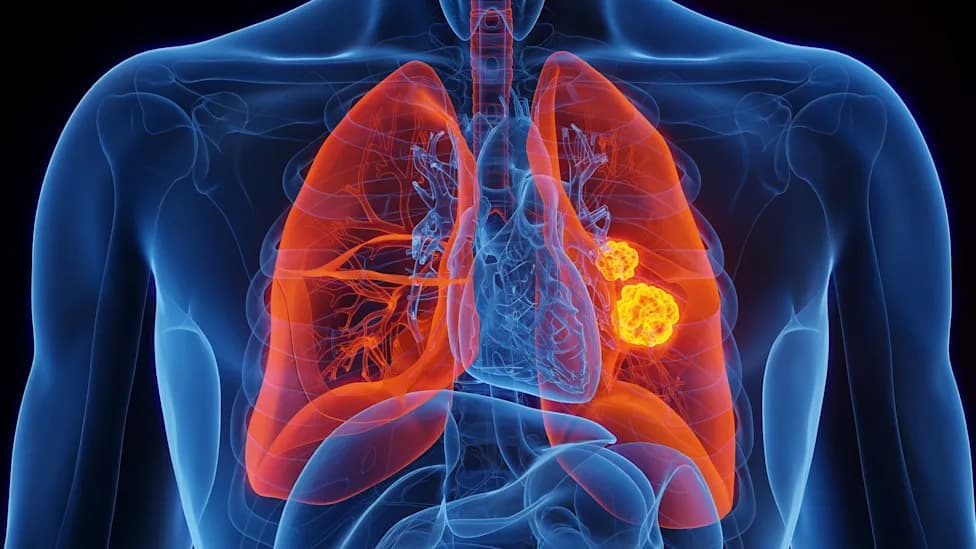
A PET imaging study from the Centre for Addiction and Mental Health and the University of Toronto found reduced VMAT2—a marker of dopamine nerve terminals—across the striatum in 24 long COVID patients compared with 24 controls. Reductions ranged from about 16% in the dorsal putamen to roughly 20% in the ventral striatum and closely correlated with apathy, memory problems and slowed movement. The results build on earlier evidence of inflammation in the same regions and suggest possible pathways for targeted treatments, including a planned trial of a dopamine-related drug. Findings are promising but need replication in larger cohorts.
Imaging Study Links Long COVID Brain Fog to Loss of Dopamine Nerve Terminals

Scientists found a link between damaged dopamine nerves and those experiencing symptoms of Long Covid.
Researchers at the Centre for Addiction and Mental Health and the University of Toronto report measurable reductions in dopamine-related nerve terminals in people with long COVID, offering a biological explanation for symptoms such as brain fog, apathy and slowed movement.
What the Study Did
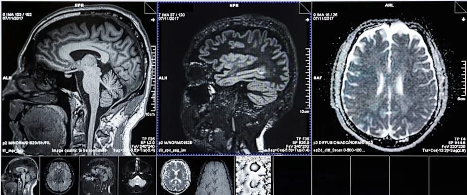
Led by psychiatry professor Jeffrey Meyer, the team used positron emission tomography (PET) to measure VMAT2, a protein located on nerve terminals that package and release dopamine. They compared 24 people with long COVID to 24 healthy controls who had experienced only mild or moderate initial SARS-CoV-2 infections.
Main Findings
The investigators found a significant reduction in VMAT2 across the striatum, the brain region involved in movement, motivation and memory. Reductions ranged from about 16% in the dorsal putamen (linked to planning and motor control) to roughly 20% in the ventral striatum (closely tied to motivation and apathy). Lower VMAT2 measures in specific striatal subregions correlated with particular symptoms: ventral striatum with apathy and memory problems, dorsal putamen with slowed movement, and a third subregion with reduced motivation.
“By showing that there are reductions in the nerves that release dopamine in these areas, and then showing the relationship of them to the symptoms, that's two pieces of information that makes a strong connection to long COVID,” Meyer told Fortune.
How This Fits With Prior Work
The new findings build on a 2023 study from the same group that detected elevated inflammation in these same brain regions. The researchers suggest two plausible, nonexclusive explanations: inflammation could damage the terminals of dopamine-releasing nerves, or primary nerve injury could provoke the inflammation.
Implications and Next Steps
Importantly, the PET marker (VMAT2) is interpreted as indicating a reduction in the density of dopamine-releasing nerve terminals rather than a temporary drop in dopamine signaling. Whether these terminal losses are reversible remains uncertain. Meyer noted some patients might regain function if terminals regrow or new connections form—possibly aided by exercise or activities that engage these brain regions—while others may require targeted medical treatment if ongoing inflammation blocks recovery.
The team is preparing a clinical trial to repurpose a dopamine-related medication known to cross into the brain; anecdotal, striking improvements have been reported in a few patients. However, funding for a full-scale trial has narrowly missed review twice, delaying larger confirmatory studies.
Limitations
Sample size was modest (24 patients versus 24 controls), so findings require replication in larger, diverse cohorts. The study shows strong associations between reduced VMAT2 and symptoms but does not by itself prove direct causation. Further work should clarify mechanisms, long-term outcomes, and which patients are most likely to benefit from dopamine-targeted therapies.
Bottom Line
This imaging study provides a tangible biological marker linked to the characteristic cognitive and motivational symptoms of long COVID and opens the door to targeted clinical trials and diagnostic approaches. If validated, the findings could shift how clinicians understand, diagnose and treat post-COVID neurological symptoms.
Help us improve.