Phase III INBUILD analyses show Brainomix’s e‑Lung can detect progression and treatment response in PPF. In 474 patients, e‑Lung and a UCLA research algorithm both identified statistically significant changes with nintedanib—TDE at weeks 24 and 52, and RVS/WRVS at week 24. Higher baseline TDE correlated with greater 52‑week FVC decline, suggesting quantitative CT can complement FVC as a trial biomarker and support earlier, more sensitive measures of lung‑structural change.
AI Imaging Breakthrough: Brainomix’s e‑Lung Predicts Progression and Detects Treatment Response in Phase III PPF Trial

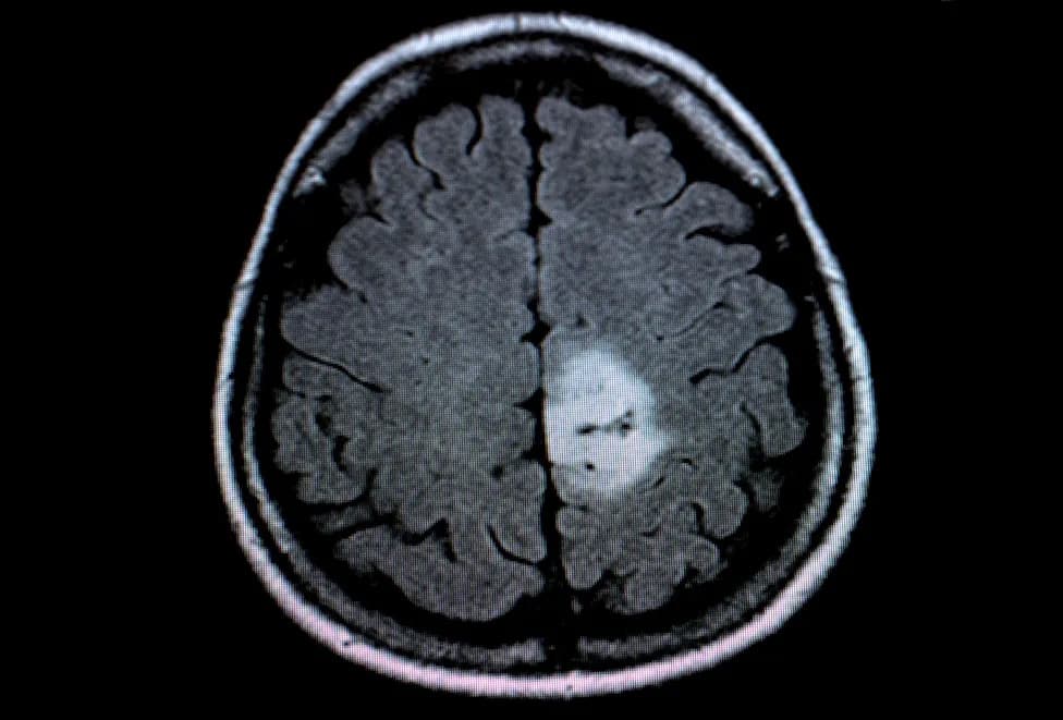
Brainomix's e-Lung was proven sensitive to the effects of antifibrotic treatment. Credit: Marko Aliaksandr / Shutterstock.com.
Brainomix today released Phase III analyses from the INBUILD trial (NCT02999178) showing that its AI-driven quantitative CT platform, e‑Lung, can both forecast disease trajectory and measure response to antifibrotic therapy in patients with progressive pulmonary fibrosis (PPF).
Study and Methods
Analysts compared CT-derived metrics generated by Brainomix’s e‑Lung software with a research algorithm from the University of California, Los Angeles (UCLA) across 474 patients enrolled in INBUILD. The goal was to evaluate whether quantitative imaging biomarkers could sensitively detect structural lung changes and relate those changes to functional outcomes over 52 weeks.
Key Findings
Both analytic approaches consistently and sensitively measured the effects of the antifibrotic agent nintedanib. e‑Lung–derived measures showed statistically significant changes in total disease extent (TDE) at weeks 24 and 52, and in reticulovascular score (RVS) and weighted reticulovascular score (WRVS) at week 24. Higher baseline TDE and other quantitative CT measures were associated with a faster decline in forced vital capacity (FVC) over 52 weeks, linking imaging‑derived disease burden with an important functional endpoint.
Implications
Brainomix and study authors suggest that quantitative CT can complement traditional endpoints like FVC by providing earlier or more sensitive signals of structural change. Such imaging biomarkers could refine clinical‑trial endpoints, improve patient stratification, and potentially shorten timelines for evaluating novel therapies for interstitial lung diseases including PPF.
Anand Devaraj, MD, Medical Director at Brainomix, said the findings support the continued development and incorporation of quantitative CT as an objective biomarker for clinical trials and that the same tools could be adapted for routine clinical monitoring of individual patients.
Perspectives From Industry
Susanne Stowasser, INBUILD co‑author and head of pulmonology/rheumatology clinical development at Boehringer Ingelheim, noted that detecting meaningful structural changes in the lung strengthens evidence for antifibrotic therapies and can inform the design of future studies.
Adoption Challenges and Market Context
The report highlights that while AI is increasingly used to triage and quantify imaging, implementation raises governance, integration and regulatory considerations that some healthcare systems still need to address. Despite these hurdles, market analysis from GlobalData projects the healthcare AI segment could reach approximately $57.4bn by 2029, underscoring strong commercial interest in imaging AI solutions.
Bottom Line
The INBUILD imaging analyses support the value of quantitative CT as a complementary tool in PPF research and potentially clinical care, demonstrating that AI‑driven imaging biomarkers like e‑Lung can detect treatment effects and predict functional decline over one year.
Trial: INBUILD (NCT02999178). Patient Cohort: 474 PPF patients. Intervention Highlighted: Nintedanib.
Help us improve.