MIT's RamanOmics pairs Raman microscopy with single-cell and spatial transcriptomics to detect biochemical fingerprints of senescent cells without destroying tissue. Tested in lung and skin from 2‑month and 26‑month mice, the platform found tissue-specific aging programs and a recurring lipid-associated Raman peak at 1,131–1,135 cm⁻¹ in p21-positive senescent cells. A random-forest classifier combining Raman and gene features produced multimodal barcodes that improved detection, though imaging speed and human validation remain major hurdles.
MIT's RamanOmics Uses Light to Detect Senescent Cells Non‑Destructively
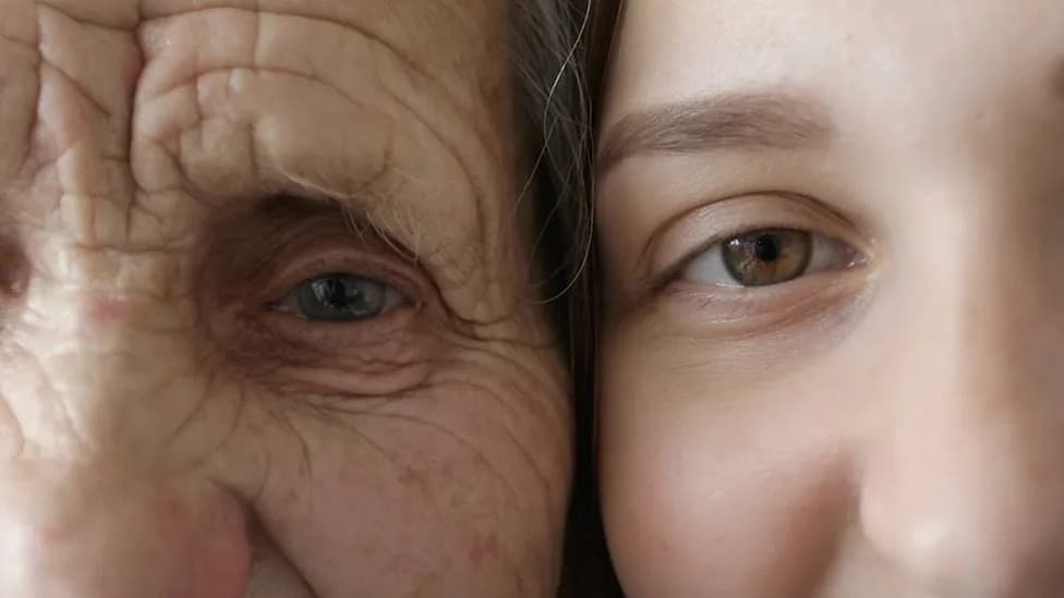
MIT researchers use Raman microscopy and gene data to identify biochemical barcodes of senescent cells without destroying tissue. (CREDIT: Shutterstock)
MIT researchers combined Raman microscopy with single-cell and spatial transcriptomics to read biochemical fingerprints of senescent cells without destroying tissue. Their multimodal platform, RamanOmics, identifies recurrent spectral features — notably a lipid-associated Raman peak — and pairs them with gene-expression data to create machine-learning “barcodes” that distinguish senescent from nonsenescent cells in intact mouse lung and skin.
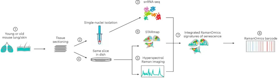
RamanOmics workflow and dynamics of gene regulation across ages and tissues. (CREDIT: Ke Zhang et al, Nature Aging)
Published in Nature Aging, the study demonstrates that optical biochemical signatures complement transcriptomic profiles and could, with further development and validation, enable nondestructive imaging tools to locate senescent cells in living tissue.
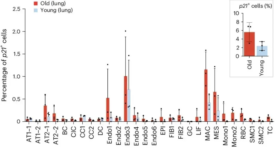
Cell-type-specific senescence programs revealed by differential expression profiling in aging lung and skin. (CREDIT: Ke Zhang et al, Nature Aging)
Why Nondestructive Detection Matters
Senescent cells stop dividing but remain metabolically active and can drive inflammation, fibrosis, and age-related disease when they accumulate. Conventional markers (for example, p16 and p21) and common laboratory techniques often require fixation, staining, or tissue dissociation, which prevents repeated or follow-up analyses on the same tissue. A nondestructive optical approach would let researchers and clinicians locate, monitor, and potentially re-sample the same tissue while preserving spatial context.
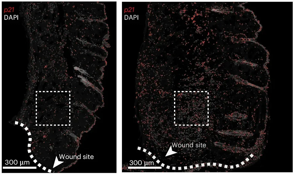
Validation of molecular and biochemical signatures in a mouse skin wound-healing model. (CREDIT: Ke Zhang et al, Nature Aging)
Methods: Multimodal, Spatial, Single-Cell Data
The team analyzed lung and skin samples from young (2-month-old) and old (26-month-old) mice. They combined hyperspectral Raman imaging with single-nucleus RNA sequencing and STARmap spatial transcriptomics to link biochemical signals to gene-expression states while preserving each cell’s position within the tissue.
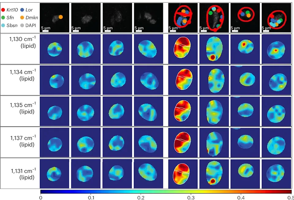
Representative Raman intensity maps of senescence-associated Raman peaks overlaid with spatial distribution of senescence-enriched differentiation genes. (CREDIT: Ke Zhang et al, Nature Aging)
Key Findings
RamanOmics revealed that aging does not produce a single universal program across organs. Aged lung tissue showed increased immune activation, inflammation, and extracellular matrix remodeling, while aged skin exhibited metabolic and structural shifts involving keratinocytes, fibroblasts, and collagen.
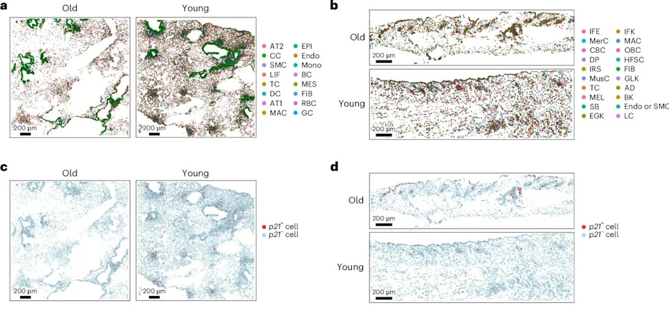
Spatial transcriptomic landscape of senescent cells in mouse lung and skin at different ages. (CREDIT: Ke Zhang et al, Nature Aging)
Senescent cells varied by tissue and age: older lung senescent cells carried signatures aligned with fibrosis and inflammation, whereas younger senescent cells showed stronger DNA-damage-repair and regenerative signals. These results support the idea of multiple senescent "senotypes" shaped by tissue context and age.
On the biochemical side, researchers detected dozens to hundreds of spectral differences between senescent and nonsenescent cells. One of the most consistent signals was a lipid-associated Raman peak near 1,131–1,135 cm⁻¹, elevated in p21-positive senescent cells across both lung and skin. Other spectral changes mapped to proteins, nucleic acids, sugars and collagen and were often tissue specific.
From Spectra to Machine-Learning Barcodes
The authors trained a random-forest classifier that combined selected Raman peaks with transcriptomic features. Integrating optical and gene-expression data improved senescent-cell classification versus transcriptomics alone. The most informative features were organized into multimodal "barcodes" intended to guide future, faster optical screening without routine RNA sequencing.
Limitations And Next Steps
The work is preclinical and performed in mouse tissues. Human tissues are more heterogeneous, and senescent states differ with disease, organ, age, and inducing stressors, so extensive validation across human samples is required. Practical limitations include imaging speed: the current system requires roughly 30 hours to scan ~1 mm². The researchers are developing faster instruments tuned to the most informative wavelengths to make clinical translation feasible.
Conclusion
The study establishes that senescent cells leave measurable biochemical fingerprints that can be read with light-based methods. While translation to humans and practical clinical imaging remain future steps, RamanOmics points to a promising nondestructive path for locating and tracking senescent cells in intact tissue — a capability that would be valuable for basic research and for testing senolytic or other interventions.
Reference: Ke Zhang et al., RamanOmics, Nature Aging (authors include Jeon Woong Kang and Salvatore Sorrentino).
Help us improve.