Researchers at the University of Rome Tor Vergata reviewed over 100 studies to map how genetic risk, TG2 enzyme activity, T cells, intraepithelial lymphocytes, stromal cells, the gut microbiome, and type I interferons interact to amplify gluten-triggered intestinal damage. The review argues that multiple environmental and immunological 'amplifiers' are required—beyond genetic susceptibility—to produce the tissue destruction seen in celiac disease. Improved disease models and preclinical testing are needed to determine the sequence of pathogenic events and to evaluate therapies that could complement a gluten-free diet.
How Immune and Microbial Signals Amplify Gluten Damage in Celiac Disease — A 100+ Study Review
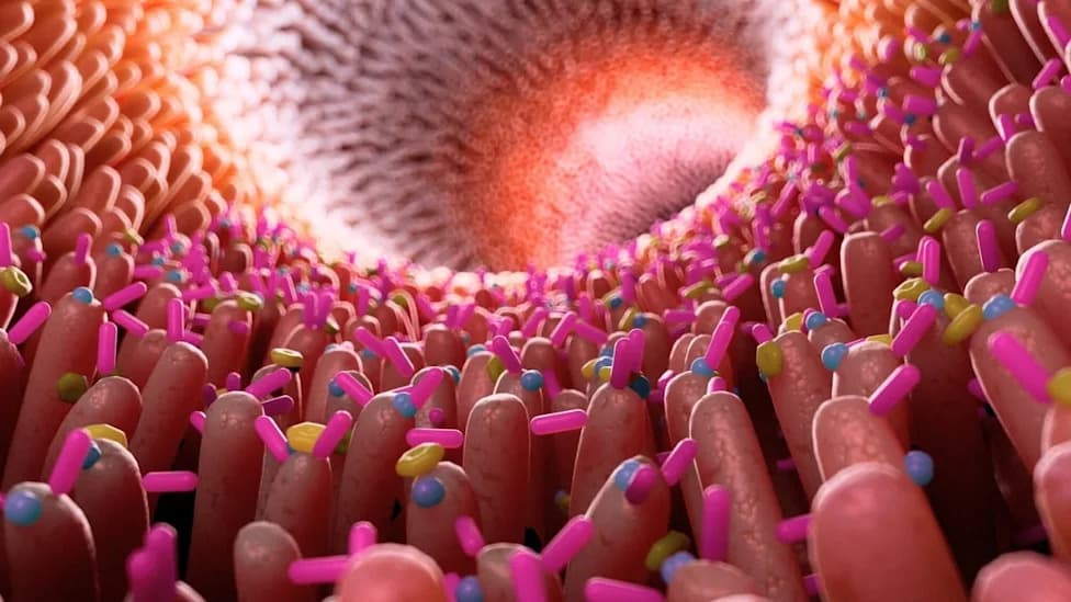
(Science Photo Library/Canva)
Celiac disease is a lifelong autoimmune condition in which digestion of gluten triggers an immune assault on the small intestine. Patients must avoid wheat, barley, and rye to prevent intestinal injury and symptoms such as bloating, pain, diarrhea, constipation, and vomiting.
Researchers at the University of Rome Tor Vergata synthesized findings from more than 100 studies in a comprehensive review published in Frontiers in Immunology. Their goal was to map how genetic risk factors are converted into the tissue damage characteristic of celiac disease by a set of amplifying mechanisms.
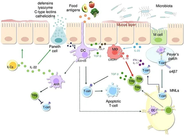
A multitude of factors contribute to a healthy gut bacteria balance. (Marafini et al.,Front. Immunol., 2026)
Key Molecular and Cellular Steps
At the molecular level, the enzyme tissue transglutaminase 2 (TG2) modifies gluten peptides (deamidation), and in people with particular HLA-associated gene variants these modified peptides become strongly immunogenic. Those peptides are presented to CD4+ T cells, which initiate an inflammatory cascade.
'Under normal conditions, TG2 is mostly inactive in the intestine. Nevertheless, inflammatory stimuli or cellular stress can increase the production and enzymatic activity of TG2, thus escalating immune responses to gluten.'
Yet T cells are not the whole story. The inflammatory milieu activates intraepithelial lymphocytes (IELs), a population of cytotoxic immune cells that directly kill epithelial cells lining the gut. This cytotoxic activity contributes to villous atrophy and impaired intestinal function.
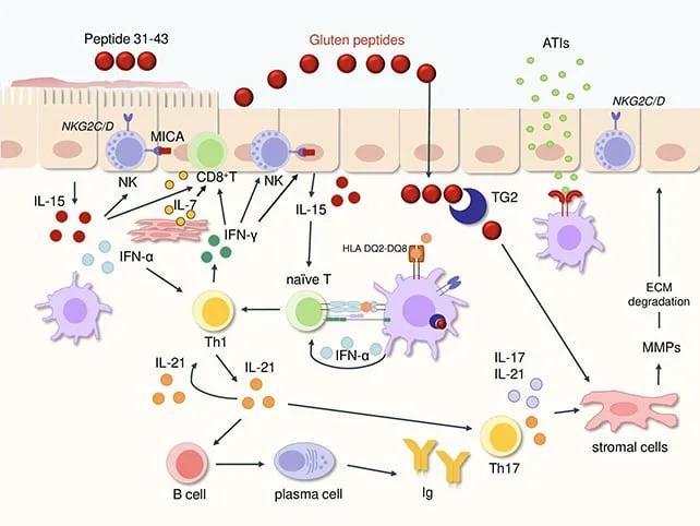
A multitude of disruptive problems occur in celiac disease. (Marafini et al.,Front. Immunol., 2026)
Stromal Cells and Additional Amplifiers
The review highlights newer evidence that stromal (structural) cells in the intestinal wall actively communicate with immune cells to sustain inflammation, rather than acting as passive scaffolding. Other amplifiers identified include variations in the gut microbiome, non-gluten wheat proteins, and antiviral-like signals such as type I interferons. Each of these factors can increase cellular stress, enhance TG2 activation, or otherwise boost immune responsiveness to gluten.
Importantly, the authors emphasize that genetic susceptibility alone is usually not sufficient to trigger full-blown disease. Instead, an interplay of genetic, environmental, microbial, and immune factors appears necessary to convert gluten exposure into sustained intestinal injury.

Subscribe to ScienceAlert's free fact-checked newsletter
Clinical Implications and Next Steps
The synthesis supports earlier work suggesting the epithelium itself is an active participant in pathogenesis. Better experimental models that reproduce the complexity of these interactions will be essential to determine the order of pathogenic events and to test new therapies. Such treatments could eventually complement or reduce reliance on strict lifelong adherence to a gluten-free diet.
Approximately 1 in 100 people worldwide may have celiac disease, making these advances broadly relevant. The review calls for continued research into microbiome contributions, TG2 regulation, stromal–immune interactions, and antiviral signaling pathways to identify candidate targets for intervention.
Source: Marafini et al., Frontiers in Immunology. Review synthesizing data from 100+ studies on celiac disease pathogenesis.
Help us improve.