Your bones may offer clues about future brain health. A new study using routine CT scans to estimate thoracic vertebral bone density found that people with lower bone mineral density (vBMD) tended to experience faster declines in thinking ability and greater progression of certain brain abnormalities on MRI.
Study Design and Methods
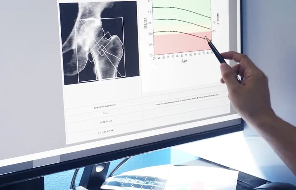
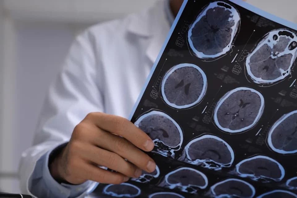
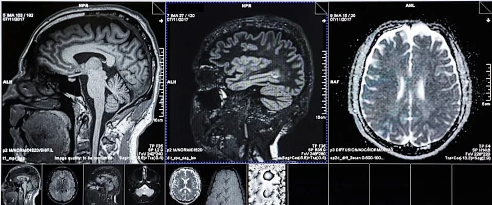
Published in Radiology, the research analyzed data from 2,086 participants drawn from an ongoing multi-ethnic cohort study. Investigators at Johns Hopkins University used an artificial intelligence algorithm to estimate volumetric bone mineral density (vBMD) in the thoracic spine from existing CT images. A subset of 715 participants also had brain MRI scans and repeated cognitive testing, enabling longitudinal comparison of bone density with brain structure and cognitive change.
Key Findings
At baseline, lower thoracic vBMD was associated with:
- Faster decline in global cognitive performance during follow-up.
- Greater accumulation of white matter hyperintensities (WMH), particularly in the corpus callosum—MRI markers linked to small-vessel damage and increased dementia risk.
- Larger declines in fractional anisotropy (a diffusion MRI measure of white matter microstructure) in the anterior limb of the internal capsule, a region involved in working memory, attention and executive function.
Subgroup Signals and Possible Mechanisms
The investigators reported exploratory evidence that some subgroups showed stronger associations: participants with diabetes exhibited a clearer link between lower vBMD and faster WMH accumulation, while the association between low bone density and loss of white matter integrity appeared somewhat stronger in women (borderline significance). The authors propose a shared vulnerability model—common metabolic, vascular, or hormonal processes (for example, dyslipidemia or menopausal changes) may affect both bone and brain—but stressed that the study does not establish causation.
"I would not want readers to come away thinking that bone loss causes brain aging, because an observational study cannot show that," said Dr. Sara Momtazmanesh, the study's lead author.
Clinical Implications and Cautions
Because the thoracic vBMD estimates were derived automatically from routine chest CTs, the finding raises the possibility that existing scans could provide additional information about patients' bone and perhaps brain health without extra imaging or radiation. Chest CTs are common in older adults, and automated vBMD extraction could one day help identify people at higher risk of faster brain aging.
However, the authors emphasize that these results need replication and validation before being used in clinical practice. "It is not a dementia test, and nothing in our study should be read as a recommendation to patients or their physicians," Dr. Momtazmanesh said.
Reference
Momtazmanesh S., et al. "Deep Learning-derived Bone Mineral Density and Longitudinal White Matter Microstructure and Cognitive Decline: Multi-Ethnic Study of Atherosclerosis." Radiology.