University of Sydney researchers developed CaAl‑DMSN, biodegradable calcium‑aluminosilicate nanoparticles that activate latent TGF‑β1 and promote rapid clotting and recruitment of bone progenitor cells. In a rat critical‑size calvarial defect model, treated defects showed ~80% greater bone volume fraction after eight weeks, and latent TGF‑β1 activation exceeded conventional alkaline methods by over tenfold. The team plans to explore pairing the nanoparticles with patient‑specific 3D‑printed scaffolds, but stresses that substantial preclinical work and careful human trials are still required before clinical use.
Biodegradable “Nanobone” Activates Body’s Repair Signals and Boosts Bone Regrowth by ~80% in Rats

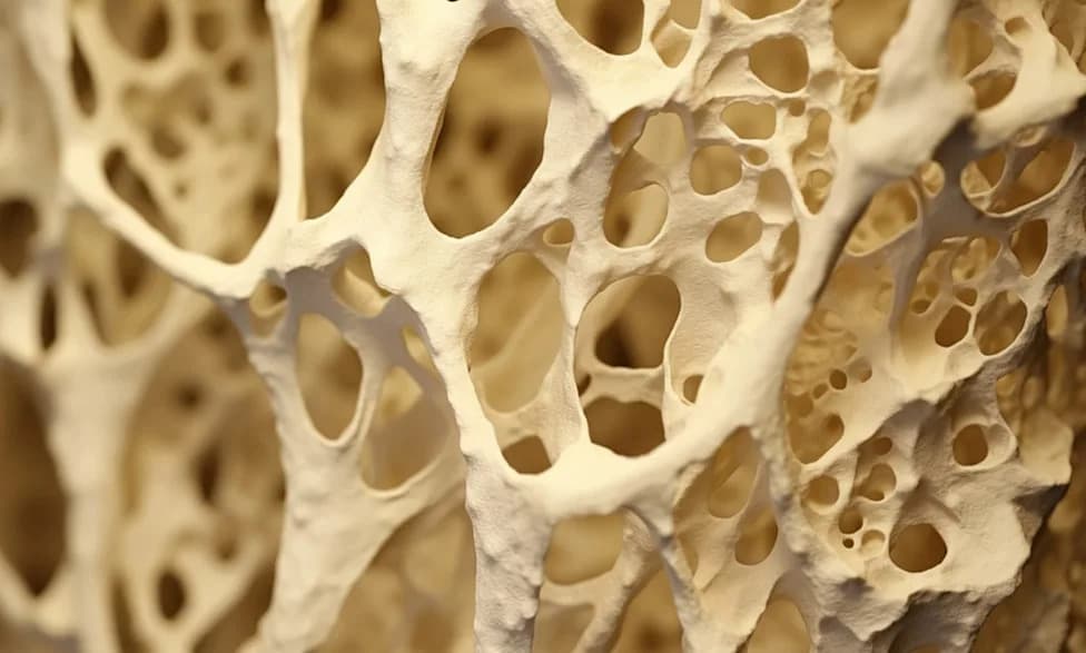
A biodegradable nanomaterial helped rats regrow substantially more bone by activating repair signals already present in the body. (CREDIT: Shutterstock)
Researchers at the University of Sydney have engineered a biodegradable nanoparticle, dubbed CaAl‑DMSN, that stimulates the body’s own repair machinery to regenerate bone. Published in ACS Nano, the preclinical work demonstrates that these calcium‑aluminosilicate, dendritic mesoporous silica nanoparticles can release active transforming growth factor‑beta 1 (TGF‑β1) from its latent complex and foster rapid early wound stabilization—without delivering external growth factors.
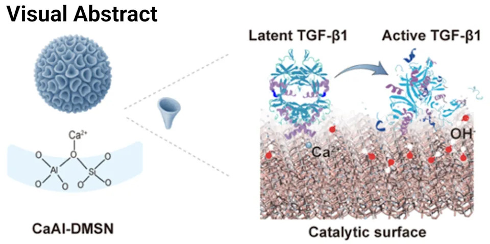
Visual abstract: A biomedical schematic showing porous calcium-aluminosilicate nanoparticles acting at a bone injury site. (CREDIT: Chun Xu et al, ACS Nano)
How the Material Works
The nanoparticles are doped with calcium and aluminum and have a radial pore architecture that creates a locally confined alkaline microenvironment. That local chemistry helps liberate active TGF‑β1 from its latent binding proteins. Laboratory assays and molecular‑dynamics simulations showed the particles activate latent TGF‑β1 at more than 10× the efficiency of conventional homogeneous alkaline treatment and appear to lower the energy barrier for growth‑factor release.
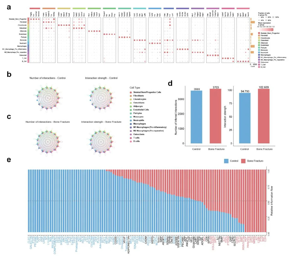
Cellular heterogeneity and global communication network changes during bone healing. (CREDIT: Chun Xu et al, ACS Nano)
Biological Effects Observed
Beyond biochemical activation, CaAl‑DMSN promotes rapid hemostasis—clot formation occurred in roughly 30 seconds in vitro—helping stabilize a wound and provide an early scaffold for repair. Cellular experiments showed increased recruitment of mesenchymal stromal cells (CD105+, CD73+). Under the nanoparticle‑induced signaling environment, signaling through the TGF‑β/Smad pathway encouraged osteogenic differentiation, directing multipotent cells toward a bone‑forming fate.
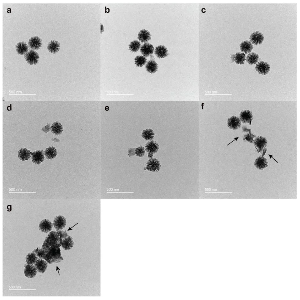
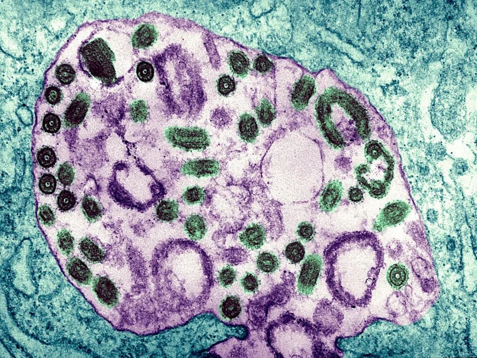
TEM images of CaAl-DMSN after incubation in PBS at 37 °C for 0 weeks (a), 1 week (b), 2 weeks (c), 3 weeks (d), 4 weeks (e), 5 weeks (f), and 6 weeks (g). (CREDIT: Chun Xu et al, ACS Nano)
Preclinical Results
In a standard preclinical model—critical‑size calvarial defects in rats (defects that do not reliably self‑repair)—treatment with CaAl‑DMSN produced approximately an 80% increase in bone volume fraction after eight weeks compared with untreated controls. The authors reported good biocompatibility in these experiments and characterized particle behavior over 0–6 weeks by transmission electron microscopy.
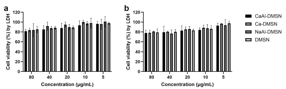
LDH cytotoxicity assay of MC3T3-E1 cells treated with different nanoparticles at 2 (a) and 6 (b) h. (CREDIT: Chun Xu et al, ACS Nano)
Potential Application and Future Directions
The team frames this strategy as a potential alternative to autologous bone grafting for alveolar cleft reconstruction in children with cleft lip and palate, where surgeons currently harvest iliac crest bone to fill jaw defects. Rather than implanting manufactured growth factors or taking bone from elsewhere in the body, CaAl‑DMSN aims to mobilize endogenous TGF‑β1 at the injury site.
Researchers are exploring integration of these nanoparticles with patient‑specific 3D‑printed scaffolds to provide both biochemical cues and a structural framework tailored to complex craniofacial geometry. If successful and safe in humans, such a resorbable material could reduce the need for a second surgical site to harvest bone.
Important Limitations and Safety Considerations
These findings are encouraging but remain preclinical. A rat calvarial model differs substantially from a growing human jaw: safety, optimal dosing, degradation kinetics, long‑term bone quality and potential effects on developing teeth must be thoroughly evaluated. Human trials have not yet begun, and substantially more preclinical testing is required before clinical application for cleft‑related jaw reconstruction.
Lead author Associate Professor Chun Xu noted: “The material activates dormant repair signals in the body, triggering a cascade of healing processes that attract bone‑forming stem cells and stimulate new bone growth.”
Next steps include expanded safety studies, optimization of particle dose and degradation behavior, long‑term assessments of regenerated bone quality, and evaluation of tooth development effects before any human testing.
Help us improve.