The Harvard team found that the mouse olfactory epithelium contains a precise, dorsoventral map of roughly 1,100 receptor expression peaks, arranged as overlapping horizontal stripes rather than random zones. Using single‑cell sequencing, spatial transcriptomics and MERFISH across ~5.5 million cells from 300+ mice, the study shows that positional transcriptional signatures and a retinoic acid gradient bias receptor choice early in development. Those epithelial positional codes also predict glomerular positions in the olfactory bulb, linking peripheral organization to central wiring and suggesting new leads for therapies to restore smell.
Harvard Team Reveals a Precise Map of Smell Receptors Running Top-to-Bottom in the Nose

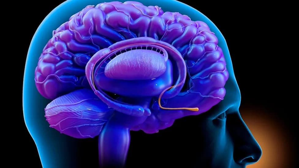
Harvard researchers mapped smell receptors in mice, showing the nose is far more organized than scientists believed. (CREDIT: Shutterstock)
For decades smell was considered the odd sense out — messy, unstructured and unlike the tidy maps seen in the eye, ear and skin. A new large-scale study led by Sandeep (Robert) Datta at Harvard Medical School shows that the olfactory epithelium in mice instead contains a precise, layered map of receptor identities that runs dorsoventrally (from the top to the bottom of the nose).
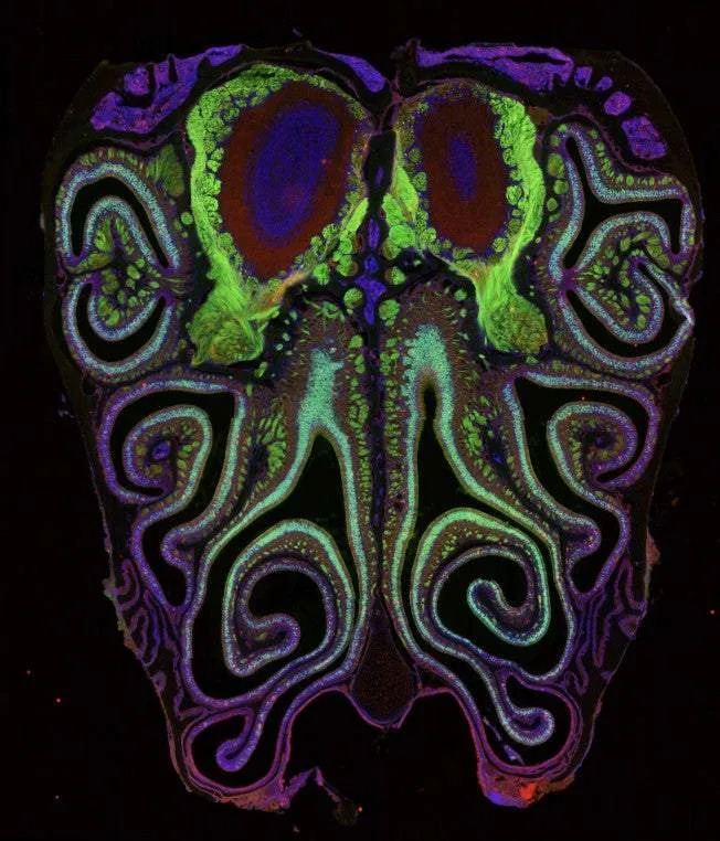
A microscope photo of a cross section of a mouse nose. The mouse was genetically modified to express green fluorescent protein in smell neurons. A small subset of dying neurons is labeled in red. (CREDIT: Datta Lab)
Key Findings
The team identified roughly 1,100 distinguishable peaks of olfactory receptor expression organized as overlapping horizontal stripes rather than as broad, random zones. These positional patterns were highly stereotyped across animals and were visible both in sequencing data and in direct spatial imaging.
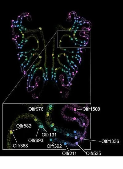
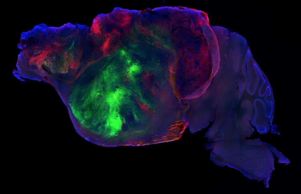
A map of the thousand types of smell receptors in the olfactory tissue of a mouse nose, labeled by a color gradient. The bottom inset shows the precise spatial positions of a tagged subset of receptors. (CREDIT: Datta Lab)
How the Study Was Done
The researchers combined single-cell RNA sequencing with spatial transcriptomics and MERFISH imaging. Altogether they profiled about 5.5 million cells from more than 300 mice, including ~2.3 million olfactory sensory neurons (OSNs). This scale allowed the group to pair each neuron's expressed receptor with its precise position in the epithelium.
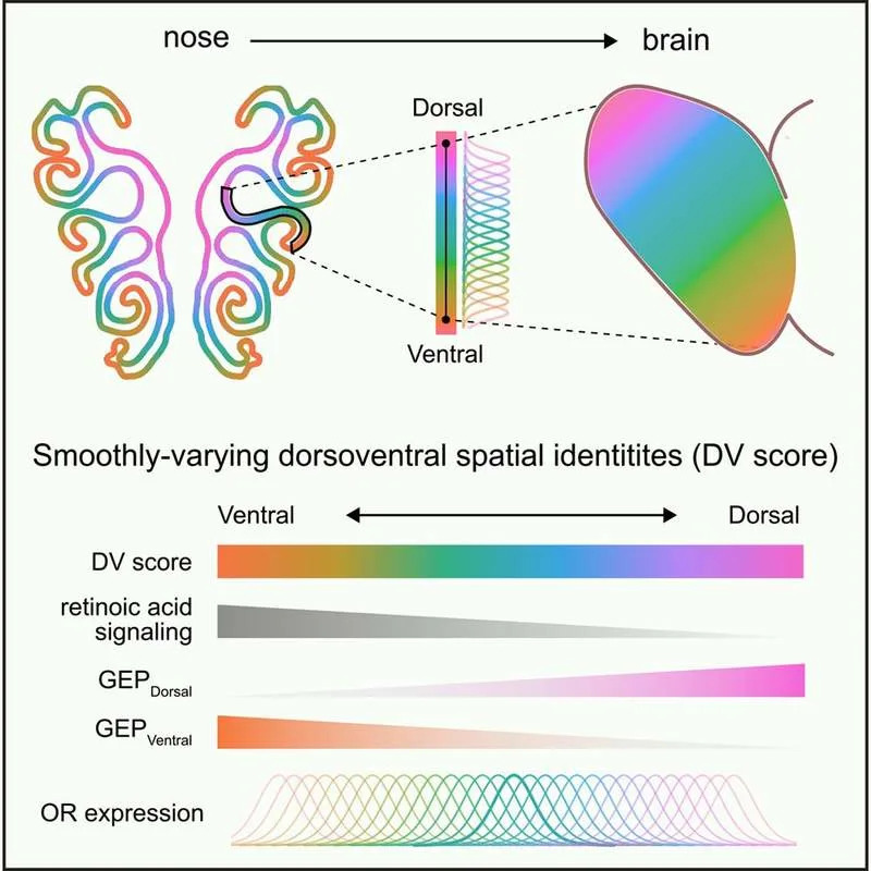
Graphical abstract. Spatial order in the olfactory system, therefore, arises from a continuously varying transcriptional code that precisely organizes the many discrete channels responsible for smell. (CREDIT: Cell)
How the Map Is Built
Evidence points to graded positional cues in the tissue. A dorsoventral gradient of retinoic acid–related signaling in the mesenchyme beneath the epithelium aligns with receptor positions above it. Olfactory stem and precursor cells carry the machinery to read that gradient: manipulating retinoic acid during regeneration shifted the map up or down the dorsoventral axis. These results indicate that positional transcriptional signatures are established early, bias receptor choice before a neuron commits to a single receptor, and produce tight but not absolute spatial distributions of receptor identity.
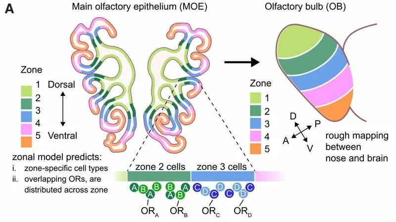
Schematic depicting the zonal model for OR choice. (CREDIT: Cell)
From Nose To Brain
OSNs expressing the same receptor converge their axons onto glomeruli in the olfactory bulb. The study found that the same dorsoventral transcriptional signatures that mark receptors in the epithelium also predict where corresponding glomeruli will form in the bulb. Coloring glomeruli by epithelial positional scores shows neighboring glomeruli share similar scores and that glomerular position predicts receptor identity with high accuracy — linking peripheral spatial codes to early central wiring.
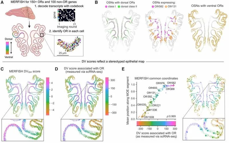
Schematic depicting multiplexed detection of ORs and non-OR genes (including DV genes) in the MOE via MERFISH, with the DV axis colored along an example epithelial segment. (CREDIT: Cell)
Limitations And Open Questions
The work was done in mice; the authors are now examining human tissue to test cross-species generality. The investigators also report only weak overall chemotopy: related receptors sometimes cluster but most odor tuning remains distributed across the epithelium. There is biological variability — ventral receptors tend to be more broadly distributed than dorsal ones, and receptor choice retains some stochasticity — so the map is precise but not rigid.
Why This Matters
“Our results bring order to a system that was previously thought to lack order, which changes conceptually how we think this works.” — Sandeep Datta
Beyond reshaping basic understanding of olfaction, the receptor map offers practical value. It provides foundational knowledge for attempts to restore smell after injury or disease — including stem-cell–based strategies or engineered interfaces — and may guide future therapies for smell loss, a condition that affects safety, taste and emotional health and gained widespread attention during the COVID‑19 pandemic.
Publication note: The findings appear in Cell; a companion study from Catherine Dulac’s lab in the same issue reached consistent conclusions.
Help us improve.