The study built a single-nucleus molecular atlas of 89,494 V1 interneuron nuclei from the mouse spinal cord and defined 14 molecular clusters. A small V1Pou6f2/Nr5a2 subgroup enriched for Piezo2 was selectively depleted when Engrailed1 (En1) was removed and was linked specifically to slower rhythmic locomotion. Broad V1 loss slowed rhythm and caused severe hindlimb hyperflexion, showing that speed and flexion are controlled by distinct V1 subsets. Core V1 identities persisted from birth to adulthood, although many genes shifted early postnatally.
Single-Cell Atlas of 89,494 Spinal Neurons Links a Tiny V1 Subgroup to Walking Speed
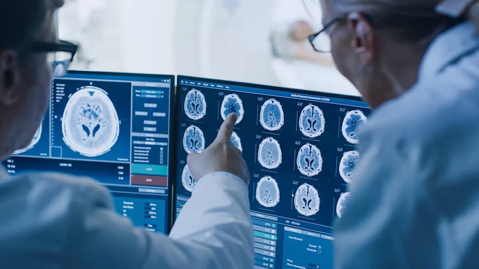
A small V1 neuron population affected locomotor rhythm without causing the limb hyperflexion seen after broader V1 loss. (CREDIT: Shutterstock)
Researchers have produced a high-resolution single-nucleus molecular atlas of V1 interneurons in the mouse spinal cord and identified a small, specialized subgroup that helps set the pace of rhythmic locomotion. The dataset — built from 89,494 V1 nuclei sampled at birth and at 14, 28 and 56 days — reveals molecular diversity within the inhibitory neurons that shape movement and points to distinct V1 populations controlling speed versus limb flexion.
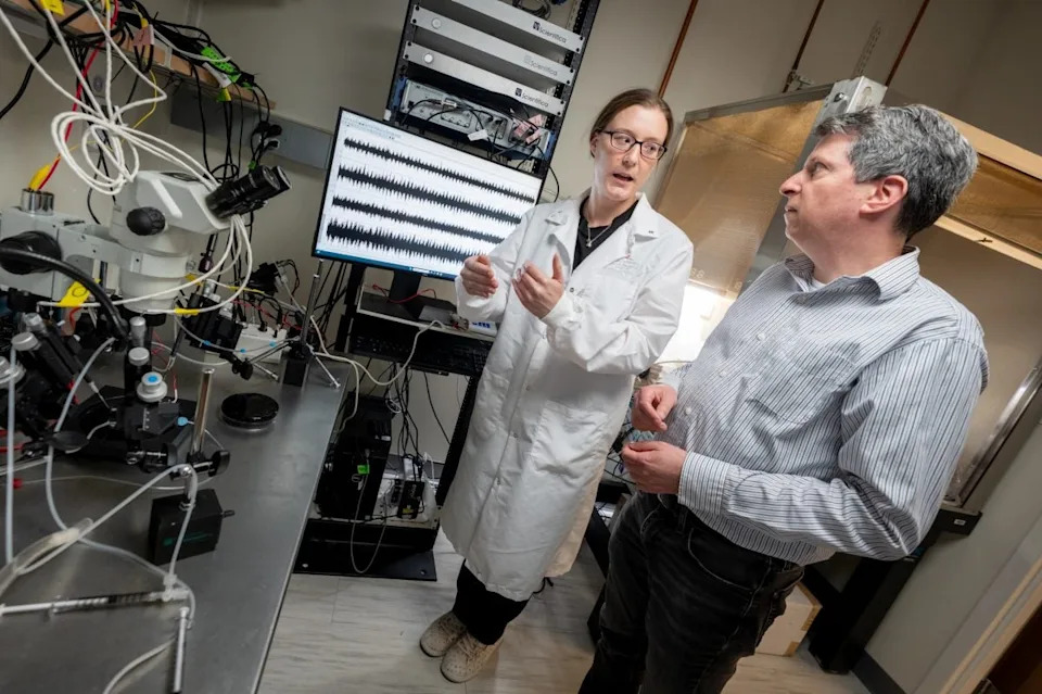
First author Alex Trevisan, PhD, and corresponding author Jay Bikoff, PhD, both of the St. Jude Department of Developmental Neurobiology. (CREDIT: St. Jude Children's Research Hospital)
How the Study Was Done
Scientists at St. Jude Children's Research Hospital used single-nucleus RNA sequencing to profile V1 interneurons, inhibitory spinal cells that integrate signals from the brain and peripheral sensory systems before influencing motor neurons. After quality control, the developmental dataset included 89,494 V1 nuclei. To probe genetic dependencies, the team also sequenced 7,553 nuclei from control and Engrailed1 (En1) knockout animals and performed anatomical and behavioral assays, including joint-angle tracking and locomotor-like rhythm measurements.
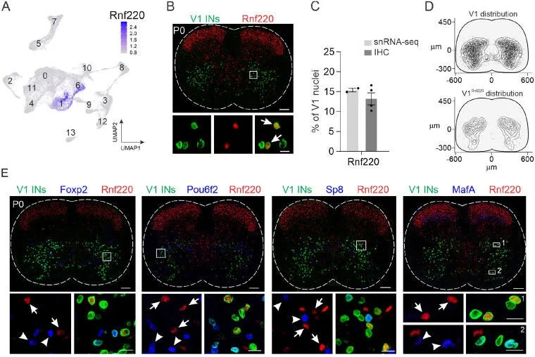
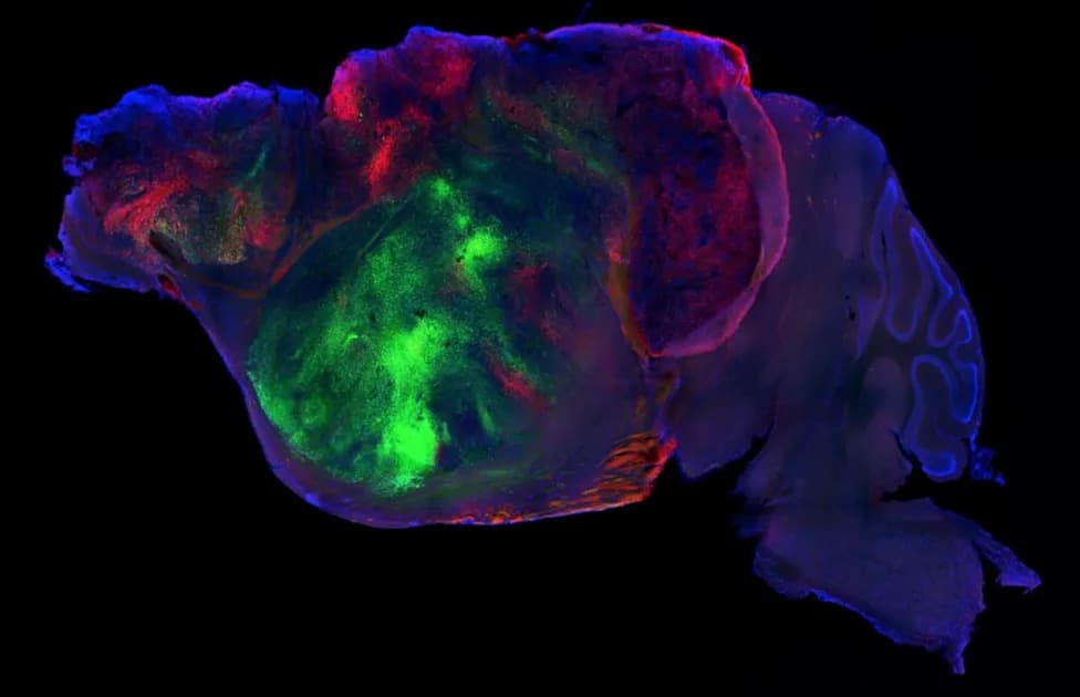
Identification of a novel V1 interneuron subset. (CREDIT: PubMed Central)
Main Findings
Fourteen Molecular Clusters: Clustering of gene-expression profiles separated V1 nuclei into 14 distinct molecular groups. The largest cluster accounted for 22.9% of cells and the smallest for 2.2%.
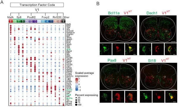
Molecular profiles of V1 interneuron subsets. (CREDIT: PubMed Central)
Five Major Clades Dominate: Four previously recognized marker-defined clades (Foxp2, Pou6f2, Sp8 and MafA) and an additional Rnf220-marked group together represent more than 95% of V1 interneurons. Only 39 of the 89,494 nuclei co-expressed markers from more than one major group, indicating clear molecular boundaries.
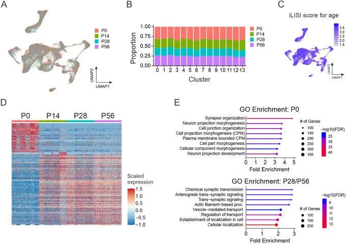
Changes in gene expression across postnatal development do not change core neuronal identity. (CREDIT: PubMed Central)
Molecular Heterogeneity Within Clades: Even within broad clades, neurons showed varied transcription-factor combinations and ion-channel repertoires. Differential expression included 13 voltage-gated potassium channel genes, 10 ionotropic glutamate receptor genes, and differences in several calcium and TRP channel genes — molecular features that likely underlie functional specialization.
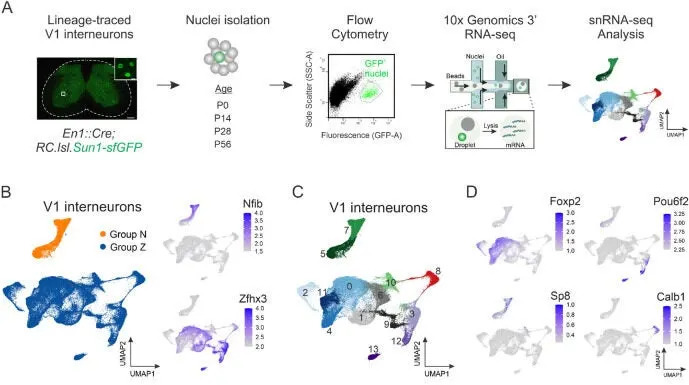
Single-nucleus transcriptomic profiling identifies V1 clades as molecularly distinct subsets. (CREDIT: PubMed Central)
Piezo2-Enriched Subgroup: A subgroup within the V1Pou6f2 clade stood out for high expression of Piezo2, a mechanosensitive receptor gene. The team validated Piezo2 expression in V1 neurons using genetically labeled mice, suggesting this subgroup may be specialized to sense mechanical signals relevant to locomotion.
Developmental Stability With Gene-Level Changes: All four ages sampled contributed across the 14 clusters in similar proportions, indicating that core V1 identities established near birth persist into adulthood. However, many genes shifted expression during postnatal maturation — especially in the first two weeks — with developmental genes (e.g., Sema6d) declining and synaptic-transmission genes (e.g., Snap25) increasing.
Genetic Perturbation Reveals Functional Specificity
Removing the transcription factor En1 produced selective changes in V1 composition without substantially changing total cell counts (control: 268 ± 9 V1 neurons per section; En1 knockout: 265 ± 7). Sequencing from control and En1-knockout tissue revealed that one cluster within the V1Pou6f2 group — marked by Nr5a2 — was nearly eliminated, falling from 6.3% of V1 cells in controls to 0.1% after En1 loss. The broader V1Pou6f2 group declined from 13.9% to 8.4%.
Behavioral Consequences: Broad ablation of V1 neurons slowed locomotor-like rhythms from roughly 0.38–0.44 Hz to ~0.13 Hz and produced severe hindlimb hyperflexion, with markedly reduced ankle and knee angles. By contrast, En1-dependent loss that selectively depleted the V1Pou6f2/Nr5a2 subgroup caused slowed rhythmic activity but did not produce hyperflexion: joint angles remained similar to controls. These results separate control of locomotor speed (implicated with the En1-dependent V1Pou6f2 subgroup) from control of limb flexion/extension (mediated by other V1 subsets).
Why This Matters
This molecular atlas provides a detailed resource to test how specialized spinal interneurons set movement parameters and respond to descending brain inputs or peripheral feedback. By identifying molecular markers and channel-expression differences, the dataset will help guide targeted experiments, and may inform strategies to restore movement after spinal cord injury or disease.
"We found that slowed locomotor speed and hyperflexion were separable, suggesting they may be controlled by different cells," said corresponding author Jay Bikoff of the St. Jude Department of Developmental Neurobiology.
Related work cited by the authors includes mapping of descending brain inputs to V1 interneurons, studies of V1 birthdates and projections, investigations of inhibitory neuron vulnerability in ALS models, and harmonized spinal-cord cell atlases that place these findings in a broader anatomical and disease context. The full dataset and analysis are available via PubMed Central.
Help us improve.