MIT researchers discovered that, when injected on axis at near‑critical power, a standard multimode fiber can self‑organize into a stable, tightly localized "pencil beam" (≈2.5 μm FWHM, 212 fs). The beam extends axial focus about tenfold while maintaining lateral resolution and reduced fluctuation, enabling 3D volumetric imaging roughly 25× faster than conventional scanning. Applied to a human blood–brain barrier model, the method captured rapid, heterogeneous transferrin uptake at subcellular resolution. Limitations include damage thresholds (~5.5 MW), a 1 MHz laser repetition cap, and remaining theoretical and sensitivity challenges.
MIT Turns Chaotic Fiber Light Into a Self‑Organized “Pencil Beam,” Enabling 25× Faster 3D Imaging of the Blood–Brain Barrier
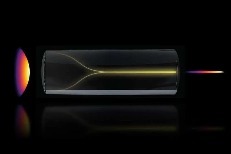
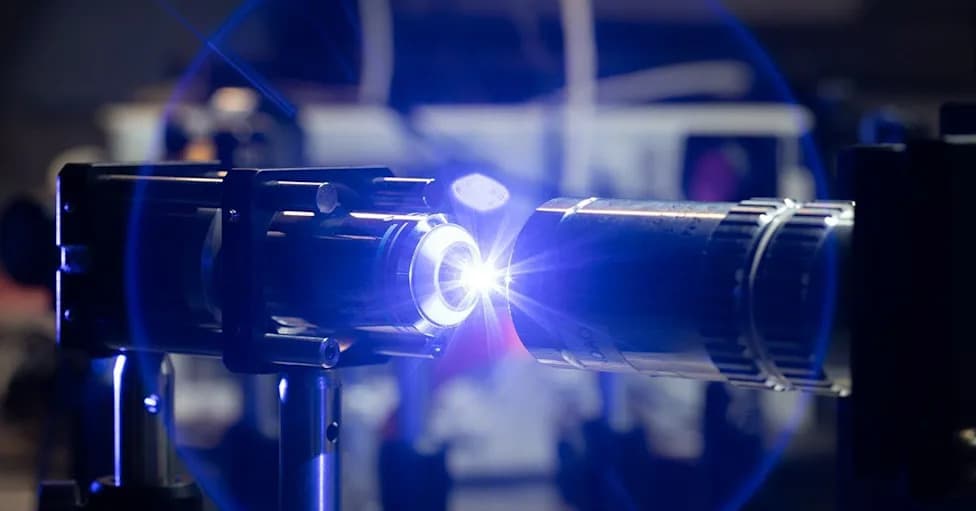
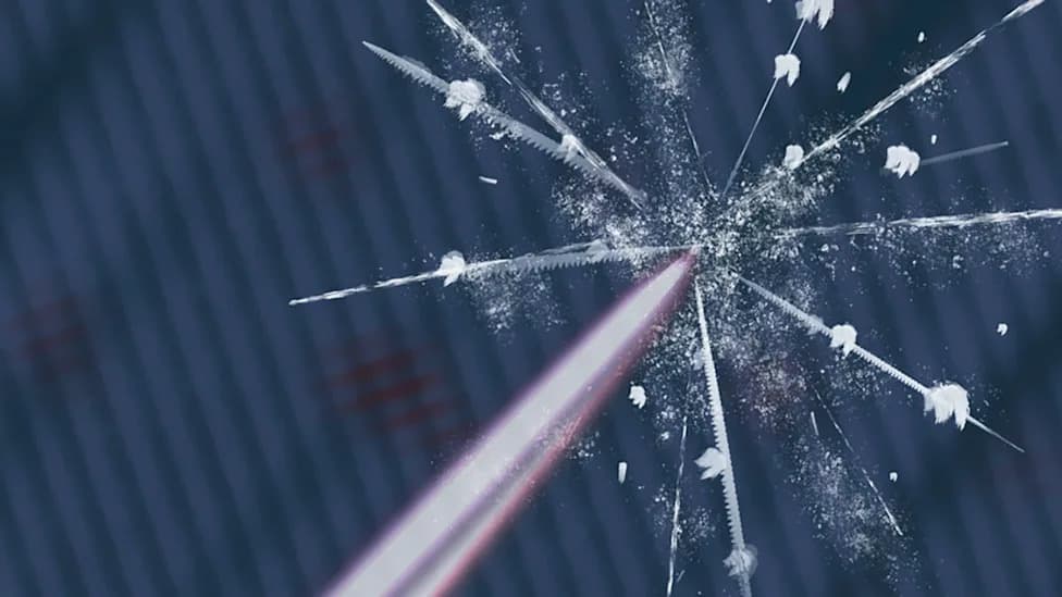
Under the right conditions, a chaotic mess of laser light can spontaneously self-organize into a highly focused “pencil beam.” This schematic shows the pencil beam formation mechanism. (CREDIT: MIT Researchers)
Researchers at MIT have demonstrated that high-power light traveling through a standard multimode optical fiber can, under precise conditions, self-organize into a tightly confined “pencil beam” instead of breaking into a noisy speckle pattern. The team reports the finding and its imaging applications in Nature Methods.
How the effect was found
While measuring damage thresholds in a 50‑micrometer‑core silica step‑index multimode fiber, graduate student Honghao Cao and colleagues observed an unexpected sharpening of the output as they pushed power close to the fiber’s limit. The effect required two strict conditions: the laser had to be injected exactly on axis (zero degrees), and the power had to be high enough for nonlinear interaction between the light and the glass.
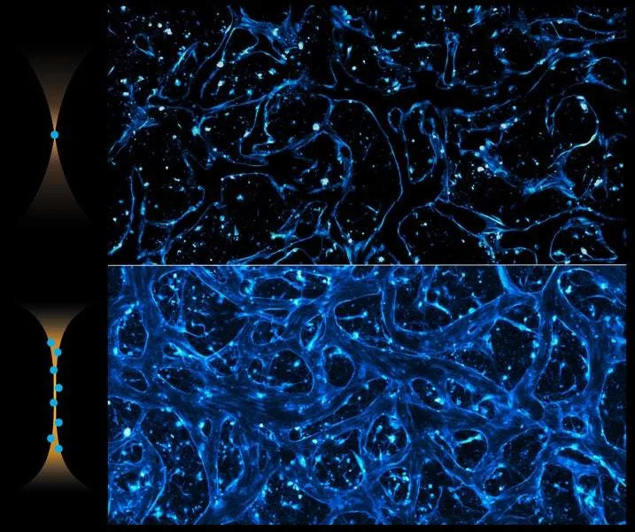
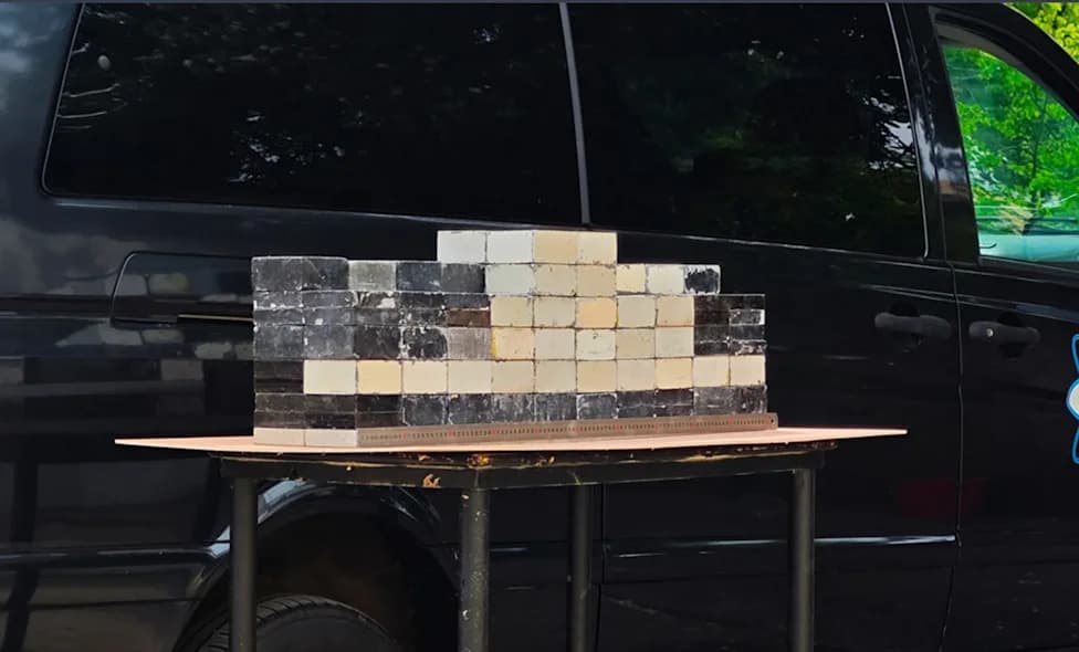
This comparison shows imaging of a blood-brain barrier model using a common Gaussian beam (top) versus the new Pencil beam method (bottom) which captures the entire volume and 3D information in a single scan. (CREDIT: MIT Researchers)
"At this critical power, the nonlinearity can counter the intrinsic disorder, creating a balance that transforms the input beam into a self‑organized pencil beam," Cao explained. Under those conditions the team measured a localized output with a full width at half maximum (FWHM) of about 2.5 μm (near the 2.3 μm diffraction limit) and pulses of roughly 212 femtoseconds.
Stability and limits
The localized state proved unusually stable: relative power fluctuations dropped by about 11 dB versus the off‑axis nonlinear output, and spatial variation under controlled bending tests decreased roughly 19‑fold. The fiber sustained the localized state for 12 hours without observable laser‑induced damage at operating settings; however, irreversible damage occurred when power reached approximately 5.5 megawatts.

The new technique enabled researchers to dynamically track how cells absorb proteins in real-time. This animation shows drug uptake (red) in a blood-brain barrier model using the pencil beam. (CREDIT: MIT Researchers)
Application to multiphoton microscopy
Multiphoton imaging typically requires a tradeoff between tight lateral resolution and limited depth of focus. Extended‑focus strategies (for example, Bessel‑like beams) increase depth but often introduce sidelobes that degrade image contrast. The MIT pencil beam keeps lateral confinement near a Gaussian reference while stretching axial focus by roughly tenfold, producing an extended depth of field without the same sidelobe penalty.
Point spread function tests with 1‑μm fluorescent beads confirmed preserved lateral resolution and ~10× axial extension. In tdTomato‑labeled mouse intestinal tissue the beam captured clear structures across an extended axial range in a single frame where a conventional Gaussian focal spot required a 50‑μm z‑stack to reveal comparable detail.
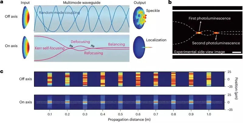
Experimental observation of nonlinear localization in a step-index MMF. (CREDIT: Nature Methods)
After repeated z‑scans (25 cycles), the speckled multimode output required ~25 mW, the localized pencil beam ~9 mW, and the Gaussian reference ~6 mW. The authors report the localized beam enabled volumetric imaging with roughly 25× shorter acquisition time and reduced photodosage relative to conventional Gaussian scanning.
Imaging the human blood–brain barrier model
The team applied the method to a microfluidic human blood–brain barrier model composed of induced pluripotent stem cell‑derived endothelial cells, pericytes, and astrocytes embedded in fibrin hydrogel. They perfused Alexa555‑conjugated transferrin through microvascular networks and acquired 3D volumes (2 mm × 2 mm × 50 μm) every minute for 50 minutes.
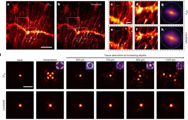
Volumetric imaging comparison of the single near-Bessel LP0n beam and the localized beam. (CREDIT: Nature Methods)
Using the self‑localized beam, imaging of a full volume took about 1 minute instead of ~25 minutes with prior settings, while preserving subcellular resolution. This speed revealed dynamic and heterogeneous uptake: endothelial cells dominated transferrin uptake and plateaued near 45 minutes, pericytes contributed under ~30% of the endothelial signal, and astrocytes showed little bulk accumulation. At single‑cell resolution, uptake varied widely—some endothelial cells internalized transferrin within minutes while neighbors remained inactive. Occasional transient "hotspots" appeared at astrocytic endfeet, and regions near pericyte coverage often correlated with reduced endothelial uptake. A competitive inhibition assay with excess unlabeled transferrin reduced endothelial signal about fivefold after 20 minutes, supporting receptor‑mediated uptake.
Implications and limitations
The technique offers a straightforward route to fast, high‑resolution volumetric imaging without custom beam‑shaping optics or adaptive correction hardware and can be generated in a standard step‑index multimode fiber, making retrofitting to existing multiphoton microscopes plausible. For preclinical drug development, this could enable time‑resolved observation of compound entry into human barrier models rather than relying solely on endpoint assays.
However, the authors note open questions and constraints: a fuller theoretical framework is needed to explain the beam’s emergence and behavior; visualizing entire transcytotic pathways remains challenging due to detection sensitivity; and current imaging speed is limited by a 1‑MHz laser repetition rate and the fiber’s energy conversion efficiency. The damage threshold (~5.5 MW) also sets an operational ceiling.
Contributors
The study was led by Honghao Cao with senior author Sixian You, and coauthors Li‑Yu Yu, Kunzan Liu, Sarah Spitz, Francesca Michela Pramotton, Federico Presutti, Zhengyu Zhang, Subhash Kulkarni (Harvard University and Beth Israel Deaconess Medical Center), and Roger Kamm (MIT). Full results are published in Nature Methods.
Help us improve.