Researchers using fMRI discovered that two-month-old infants show distinct neural responses to different object categories, suggesting earlier-than-expected category perception. The study analyzed brain scans from 130 awake two-month-olds and followed 66 of them at nine months, when category distinctions grew stronger. Lead author Cliona O’Doherty says the results reveal a more complex early interaction with the visual world, and scientists hope to connect these early brain patterns to later cognitive outcomes.
Babies See More Than We Thought: fMRI Shows Two-Month-Olds Distinguish Object Categories

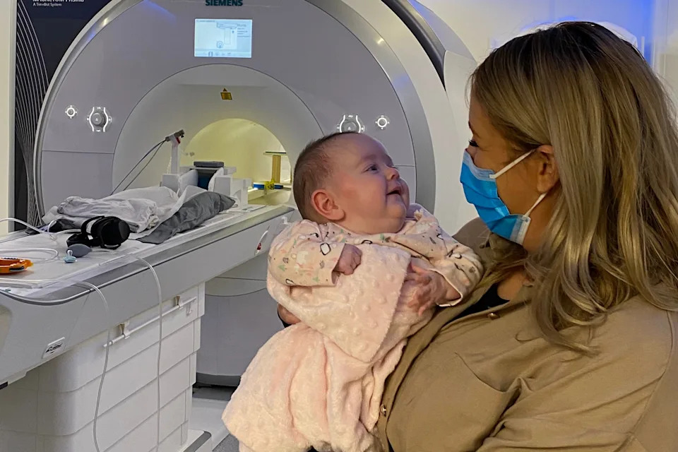
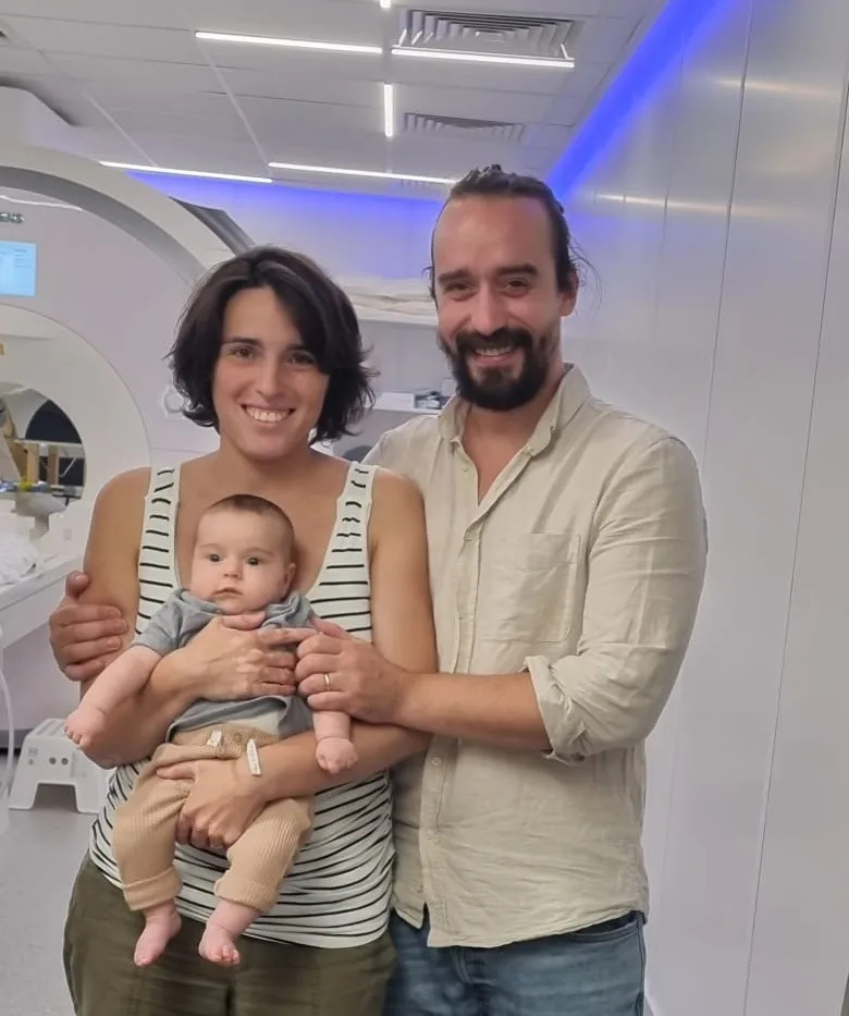
In this undated photo, Baby Blaise attends her 9-month Foundcog scan with her mother Mary at Trinity College Institute of Neuroscience in Dublin, Ireland. (Cusack Lab via AP)(ASSOCIATED PRESS)
A new brain-imaging study finds that infants as young as two months can already tell different kinds of objects apart — earlier than researchers previously believed.
Study Details
The findings, published in Nature Neuroscience, come from functional magnetic resonance imaging (fMRI) data collected while 130 two-month-old babies were awake and shown images from roughly a dozen common categories (for example, animals and trees). Researchers measured distinct patterns of neural activity when infants viewed living things versus inanimate objects, indicating category-specific responses in the infant visual system.
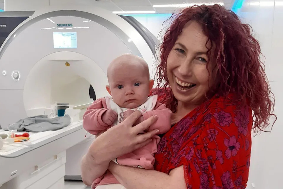
In this undated photo, baby Sadie attends her 2-month Foundcog scan with her mother Donna at Trinity College Institute of Neuroscience in Dublin, Ireland. (Cusack Lab via AP)(ASSOCIATED PRESS)
“It really tells us that infants are interacting with the world in a lot more complex of a way than we might imagine,”
— lead author Cliona O’Doherty, who led the work at Trinity College Dublin.
Why This Matters
Unlike many earlier infant studies that used looking-time as a proxy for category knowledge, this study recorded brain activity directly, giving a more precise window into early visual and cognitive organization. Past looking-time work suggested category distinctions might appear around three to four months; these fMRI results push that timing back to two months.
In this undated photo, baby Blaise attends her 2-month Foundcog scan with her mother Mary at Trinity College Institute of Neuroscience in Dublin, Ireland. (Cusack Lab via AP)(ASSOCIATED PRESS)
Many participants returned for follow-up scans at nine months; usable data were collected from 66 infants. At nine months, the neural separation between living and nonliving categories was noticeably stronger, suggesting these category representations refine rapidly over the first year.
Challenges and Practical Steps
Liuba Papeo, a neuroscientist at France’s National Center for Scientific Research, called the study’s large infant sample size “impressive and unique,” noting the technical and practical difficulties of scanning very young infants. One major challenge is keeping an infant awake, comfortable and still enough for reliable imaging.
In this undated photo, baby Maeve Truzzi-Scott attends her 2-month Foundcog scan with her mother, Dr Anna Truzzi, coauthor, and her dad, Dr. Ian Cecil Scott at Trinity College Institute of Neuroscience in Dublin, Ireland. (Cusack Lab via AP)(ASSOCIATED PRESS)
To help, the team made comfort a priority: babies reclined snugly on a bean bag in the scanner so the projected images appeared large above them. As O’Doherty put it,
“It's like IMAX for babies.”
Implications
Researchers say these early neural signatures could one day be linked to later cognitive outcomes, offering potential new ways to study typical and atypical development. The study also highlights how noninvasive imaging can reveal cognitive abilities that are not obvious from casual observation.
AP video journalist Havovi Todd contributed to this story. The Associated Press Health and Science Department receives support from the Howard Hughes Medical Institute’s Department of Science Education and the Robert Wood Johnson Foundation; the AP is solely responsible for the content.
Help us improve.