Key finding: Cryo‑EM reveals a previously unknown Z‑shaped Aβ fold in the rare Flemish APP mutation that exposes the F20 residue and likely drives vascular accumulation. The A21G mutation removes a single methyl group at residue 21, favoring this vascular‑tropic fold. Although these mutant assemblies show reduced aggregation in vitro, they form large, neurotoxic plaques in the brain and cause cerebral amyloid angiopathy with early fatal hemorrhage. Further studies will test effects on seeding, spread and toxicity.
New Z‑Shaped Amyloid Fold Explains Rare Flemish Form of Alzheimer’s

(Yana Iskayeva/Moment/Getty Images)
Alzheimer's disease takes many molecular and clinical forms. An exceptionally rare hereditary variant — caused by the Flemish amyloid‑beta precursor protein (APP) mutation — drives heavy amyloid‑beta (Aβ) accumulation around cerebral blood vessels, producing cerebral amyloid angiopathy (CAA) and often fatal brain hemorrhages by patients' late 50s.
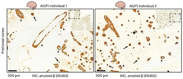
The researchers started with brain samples from two Alzheimer's patients, with amyloid-beta filaments marked with arrows. (Khaki et al.,Nat. Struct. Mol. Biol., 2026)
What the study found
An international team using high‑resolution cryogenic electron microscopy (cryo‑EM) resolved the Aβ filament structure in postmortem brain tissue from two affected individuals (one from each of the known Flemish families). The researchers discovered a previously unseen Z‑shaped Aβ fold.
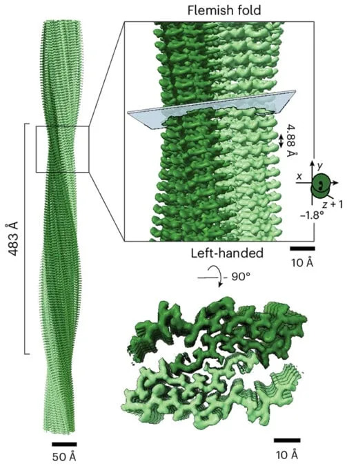
The unique way the protein folds affects the filaments it creates. (Khaki et al.,Nat. Struct. Mol. Biol., 2026)
This Z‑shaped conformation exposes the F20 amino‑acid residue on the filament surface. The team proposes that F20 exposure promotes interactions with components of the blood‑vessel wall, explaining the striking vascular targeting of Aβ in this mutation.

Subscribe to ScienceAlert's free fact-checked newsletter
The authors report that loss of a methyl group at residue 21 gives rise to an Aβ fold with vascular tropism and provides structural insight into Flemish‑type progressive dementia and cerebral hemorrhage.
How a tiny chemical change matters
The Flemish mutation (A21G) removes a single methyl group from the Aβ sequence at residue 21. That modest chemical change appears sufficient to favor formation of the distinctive Z‑fold and its vascular tropism. Previous biochemical work showed these mutant Aβ assemblies are less prone to aggregate in vitro; however, when they do form in the brain they can be as neurotoxic as typical Aβ and produce unusually large, stable plaque cores.
Why this matters
Understanding the precise shapes that Aβ can adopt — and how small sequence changes alter those shapes and tissue interactions — is critical for designing therapies that target harmful aggregates across Alzheimer’s variants. The structure reported here links a single‑atom chemical change to altered folding, vascular targeting, and a distinct clinical outcome.
Next steps
The authors say future work should test whether the Flemish fold changes seeding, propagation or neurotoxicity in cellular and animal models and whether similar structural principles underlie other hereditary or sporadic Alzheimer’s forms.
Citation: Khaki et al., Nature Structural & Molecular Biology, 2026.
Help us improve.