Researchers report the discovery of mitochondrial plaques (MPs) in neurons from Alzheimer's patients and transgenic mice. MPs are aggregates of damaged mitochondria that arise from impaired mitophagy and recruit lysosomes, leading to lysosomal dysfunction. They accumulate in neurites, often contain amyloid precursor protein (APP), appear early in disease models, and are absent from age-matched controls. The findings suggest combined therapies that target both amyloid and mitochondrial clearance could improve outcomes.
Scientists Discover Mitochondrial "Plaques" in Neurons Linked to Alzheimer's Progression
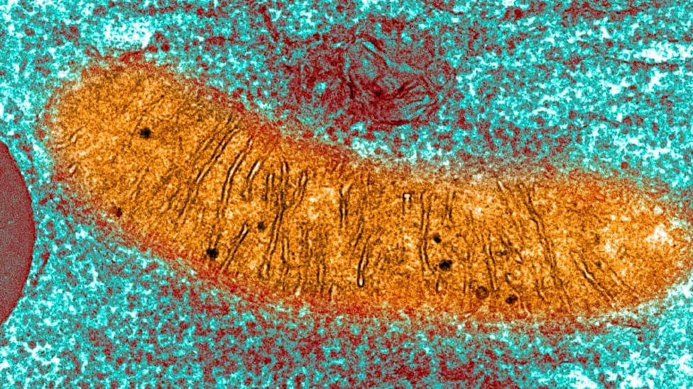
A colored image of mitochondria in a pancreas cell, via transmission electron microscopy. (Callista Images/Connect Images/Getty Images)
A multidisciplinary team publishing in Nature Neuroscience reports the discovery of previously unrecognized mitochondrial "plaques" (MPs) in the brains of people with Alzheimer's disease and in transgenic mice that model the disorder. The work highlights a new pathological feature that appears to arise from defective mitophagy — the cellular process that removes damaged mitochondria — and suggests fresh therapeutic and diagnostic avenues.
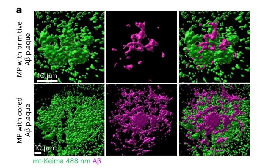
Transmission electron microscope image of the MP (green) and amyloid-beta (purple) accumulation in the brains of 50-60-week-old transgenic mice.(Dan et al.,Nat. Neurosci., 2026)
What Researchers Found
Using a genetic mitophagy reporter called Keima (a pH-sensitive fluorescent protein), the team directly tracked mitochondrial acidification and degradation in mouse brains. Keima fluoresces green in neutral mitochondria and shifts toward orange when mitochondria enter acidic compartments such as autolysosomes. Combining Keima imaging with protein assays and electron microscopy, the researchers identified aggregates of mitochondria in various states of decay: the mitochondrial plaques.

Subscribe to ScienceAlert's free fact-checked newsletter
Key Characteristics of Mitochondrial Plaques
Composition: MPs are composed of neutral and acidic mitochondria at different degradation stages and often remain trapped within acidic vesicles. The plaques frequently contain abundant amyloid precursor protein (APP), the precursor of amyloid-beta peptides.
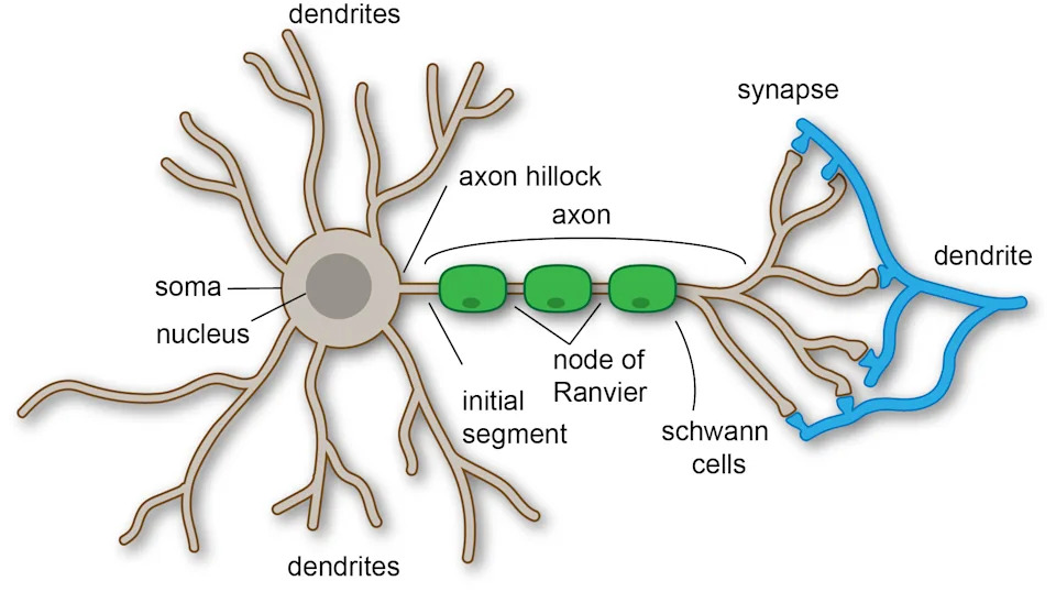
The anatomy of a neuron, including its dendrites and axon.(Curtis Neveu/Wikimedia Commons, CC BY SA 4.0)
Localization: Unlike extracellular amyloid plaques, MPs are located inside neurons — preferentially within neurites (axons and dendrites) rather than neuronal cell bodies. This positioning could impair synaptic signaling and explain persistent neurodegeneration even when extracellular amyloid is cleared.

YouTube Thumbnail
Progression: In the transgenic mouse model, MPs appeared as early as 15 weeks (an age the authors equate to early adulthood) and accumulated substantially by 50–60 weeks, with a marked rise in acidic mitochondria. Importantly, MPs were not observed in age-matched healthy control brains, in either mice or humans.
Mechanism and Cellular Consequences
The study links MP formation to impaired mitophagy and to overwhelming of the lysosomal degradation system. MPs recruit lysosomes but seem to exceed the organelles' degradative capacity, leading to lysosomal dysfunction and progressive accumulation of damaged mitochondria in neuronal processes.
Paul Robbins (University of Minnesota) says these findings 'identify mitochondrial plaques as a previously unrecognized feature of Alzheimer's disease.' Xiuli Dan, the study's first author, emphasizes that MPs act directly within neurons and may therefore be an actionable therapeutic target.
Implications for Diagnosis and Treatment
The work suggests that therapies focused solely on extracellular amyloid-beta may be insufficient because mitochondrial pathology within neurites can continue to drive degeneration. The authors propose combination strategies — for example, pairing amyloid-targeting approaches with interventions that restore mitophagy or boost lysosomal and vacuolar degradation — as a promising direction for future research.
Previous studies that induced mitophagy in Alzheimer’s mouse models showed cognitive improvements, supporting the idea that restoring mitochondrial quality control could protect neural function. The authors conclude that MPs might serve both as a new diagnostic marker and as a target for combinatorial therapeutics.
Source: Dan et al., Nature Neuroscience (2026).
Help us improve.