Italian researchers report that an intranasal spray of extracellular vesicles derived from the placental amniotic membrane preserved memory and reduced neuroinflammation in a mouse model of Alzheimer’s disease. The vesicles reached the hippocampus, lowered amyloid‑beta deposits, and increased synaptic plasticity proteins (ARC, GluA1, BDNF), while tau phosphorylation was unchanged. Complementary experiments on patient‑derived neurons showed reduced degeneration and restored plasticity markers. These promising preclinical results require further study to determine safety and efficacy in humans.
Placenta‑Derived Nasal Spray Preserves Memory and Reduces Brain Inflammation in Alzheimer’s Mice

(Tatiana Maksimova/Moment/Getty Images)
Researchers in Italy report that an experimental nasal spray made from extracellular vesicles (EVs) derived from the human placental amniotic membrane preserved cognitive performance and reduced neuroinflammation in a mouse model of Alzheimer’s disease.
What the Study Did
The study, led by cell biologist Andrea Papait at the Catholic University of the Sacred Heart, tested EVs produced by human amniotic mesenchymal stromal cells (hAMSC-EVs). Investigators formulated the vesicles into an intranasal spray and administered the treatment to transgenic mice engineered to develop Alzheimer‑like pathology. Mice received twice-weekly doses from 3 to 9 months of age (a preventative/early-intervention schedule in this model).
How the Vesicles Reached the Brain
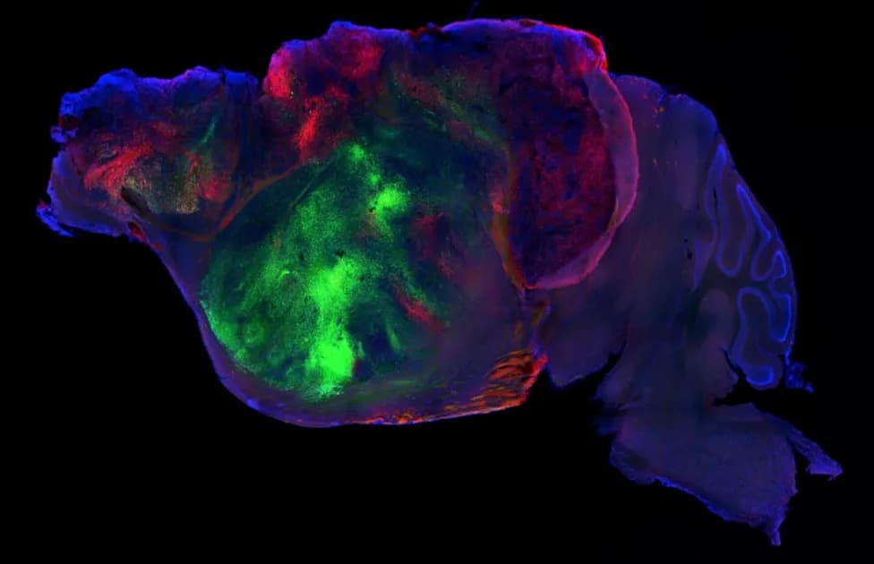
Fluorescent-tracing experiments showed that intranasally delivered hAMSC-EVs reached all regions of the hippocampus and co-localized with both neurons and microglia, supporting the intranasal route as an effective delivery method in mice.

Subscribe to ScienceAlert's free fact-checked newsletter
Main Findings
- Improved Cognition: Treated mice performed better than controls on object-recognition and spatial-memory tests, indicating preserved cognitive function.
- Reduced Amyloid‑Beta: The treatment significantly lowered amyloid‑beta deposits in the hippocampus.
- No Change in Tau Phosphorylation: Tau phosphorylation levels were not significantly affected by the EV treatment in this study.
- Lower Neuroinflammation: Markers of astrocyte and microglial activation were reduced, consistent with attenuation of neuroinflammation.
- Enhanced Plasticity Markers: Levels of synaptic and plasticity-related proteins such as ARC, GluA1, and BDNF were higher in treated animals.
Complementary Human‑Cell Experiment
In an in vitro arm, researchers reprogrammed skin cells from people with sporadic Alzheimer’s disease into induced pluripotent stem cells, differentiated them into neurons, and exposed these neurons to hAMSC-EVs. The EV treatment reduced neuronal deterioration and restored healthier expression of plasticity-related proteins, supporting translational relevance to human cells.
Proposed Mechanism
The authors suggest hAMSC-EVs modulate the inflammatory microenvironment—reshaping microglial phenotype and cytokine profiles—to promote neuronal integrity and synaptic function, rather than producing broad immunosuppression.
Limitations and Next Steps
These are preclinical results. The effects were demonstrated in a mouse model and in cultured human-derived neurons, but safety, dosing, biodistribution, and efficacy in humans remain unknown. The treatment reduced amyloid‑beta but did not affect tau phosphorylation in this study; the long-term benefits and potential side effects must be evaluated in further preclinical work and clinical trials. The researchers call for additional studies to clarify molecular mechanisms and to assess translational potential.
"These are preclinical results that require further validation in humans and do not yet represent an available therapy for Alzheimer's disease," senior researcher Claudio Grassi said. The study is published in Translational Neurodegeneration.
Help us improve.