Researchers at Xiangya Hospital combined imaging and single‑cell RNA sequencing of nearly 90,000 cells to produce a detailed map of tendon healing. They found that stalled tendon stem/progenitor cells, senescent tendon cells, macrophages reprogrammed by SOX9, and vascular cell changes actively drive collagen‑rich scarring. Two signaling pathways—osteopontin and TGF‑β—amplify fibrosis; blocking both reduced scar density in lab and animal tests. These results point to new adjunctive drugs and regenerative strategies to improve outcomes after rotator cuff repair.
New Single-Cell Map Reveals Why Rotator Cuff Tears Heal With Scar — And How We Might Stop It
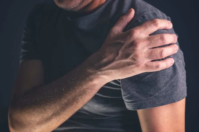
A new study shows how tendon cells and immune cells drive scar formation after rotator cuff tears, pointing to new treatments. (CREDIT: Shutterstock)
Shoulder pain often continues long after a rotator cuff tear looks healed on scans. Even after successful surgery, many patients experience persistent weakness, reduced range of motion and a high risk of re‑tear. New research from Xiangya Hospital, Central South University, offers a detailed cellular explanation for why adult tendons heal with scar tissue instead of regenerating healthy tendon.
Using advanced imaging and single‑cell RNA sequencing, the team analyzed nearly 90,000 individual cells from human tendon samples at multiple stages of healing, producing one of the most comprehensive cellular atlases of tendon repair to date. Their findings, published in Bone Research, show that fibrosis (scar formation) is an active, coordinated process driven by multiple cell types and specific molecular signals rather than a random failure of repair.
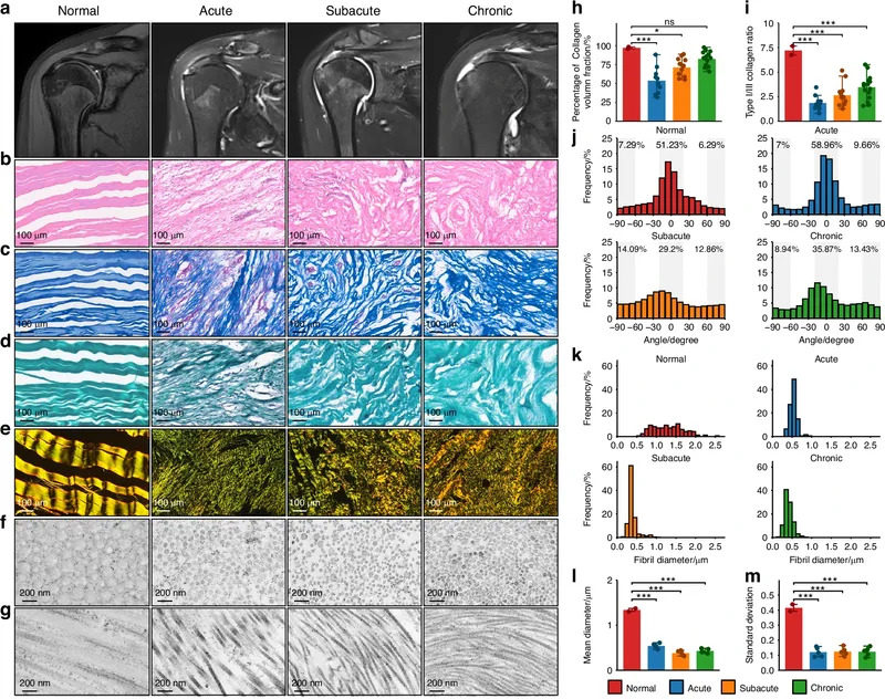
Scar formation after human rotator cuff injury. a MRI imaging of normal and different stages (acute, subacute, and chronic) of human RCT. (CREDIT: Bone Research)
Structural Changes: Collagen Disorganization and Weakening
Under the microscope, injured tendons showed early and persistent changes: normally parallel, thick type I collagen fibers became disorganized, thinner and replaced in part by type III collagen. These structural shifts reduce the tendon’s ability to bear mechanical load and help explain lingering weakness and re‑injury risk even months after repair.
Cells That Should Regenerate — But Don’t
The investigators found that tendon stem and progenitor cells, which would normally mature into functional tendon cells, often stall in an activated state and continue producing extracellular matrix components. Instead of rebuilding organized tendon, this ongoing matrix production feeds scar accumulation.
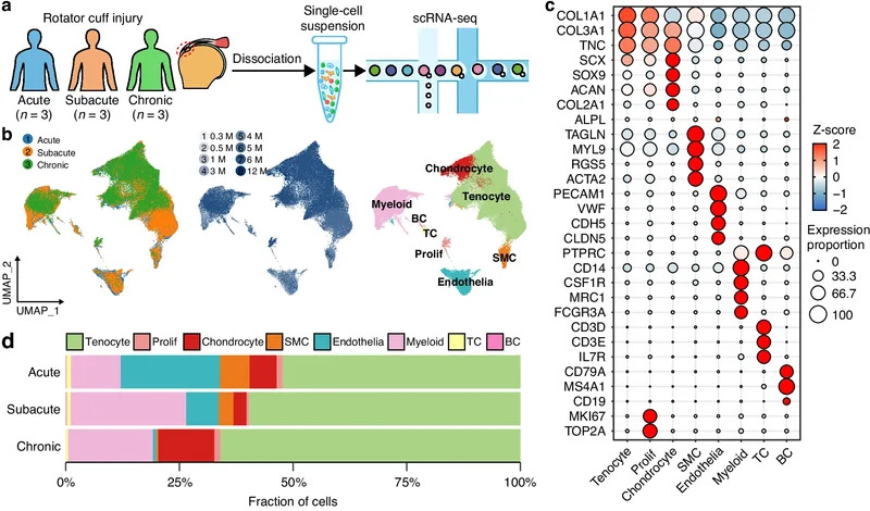
Fibrotic scar profile across stages and cell types after RCT. (CREDIT: Bone Research)
Senescence, Immune Reprogramming and Vascular Contributions
Many mature tendon cells enter a senescent state: they stop dividing and lose repair capacity but remain metabolically active and secrete signals that further stimulate collagen production. Meanwhile, immune cells—particularly macrophages—persist long after the acute inflammatory phase and undergo a striking reprogramming. Driven in part by the regulatory factor SOX9, macrophages begin producing collagen themselves and become direct contributors to the scar matrix.
Vascular cells also change identity during the transition from the acute repair phase to later remodeling. Some adopt features of matrix‑producing cells, adding another cellular source of fibrosis. Together, stalled progenitors, senescent tendon cells, reprogrammed macrophages and altered vascular cells create a self‑reinforcing, fibrotic environment.
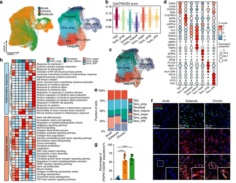
Tendon stem cell and tenocyte progenitor as the main source of ECM after RCT. a UMAP embedding of tendon cells annotated by stages and subclusters. (CREDIT: Bone Research)
Molecular Signals Behind Fibrosis
The team identified two key signaling drivers: osteopontin (produced by immune cells) and TGF‑β (a well‑known regulator of tissue repair). In cell cultures, exposure to these signals increased scar‑associated proteins; blocking them reduced collagen production. In animal models, blocking osteopontin and TGF‑β produced less dense scar tissue, with the greatest benefit when both pathways were inhibited together.
Clinical Implications and Future Directions
These results suggest fibrosis following rotator cuff injury is directed by identifiable cellular behaviors and molecular pathways that could be targeted therapeutically. Short‑term strategies might include adjunctive drugs given around the time of surgical repair to block osteopontin or TGF‑β signaling, potentially reducing scar formation and improving long‑term tendon strength and function. Longer‑term, the map of cellular states could guide regenerative approaches that steer cells back toward organized tendon reconstruction rather than scar.
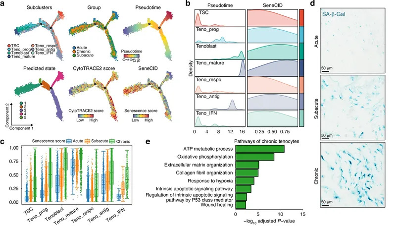
Characteristics and key regulators of senescent tenocyte after RCT. (CREDIT: Bone Research)
Study Limitations: The authors note a modest number of patient samples and that some control tissues came from a different tendon type, which may affect generalizability. Further work is needed to validate targets across larger, diverse cohorts and in clinical trials.
Because similar fibrotic programs occur in organs such as the heart, lung and liver, these findings may also inform broader anti‑fibrosis strategies. By mapping healing at single‑cell resolution, the study brings researchers closer to treatments that promote true tissue regeneration rather than imperfect scar repair.
Research Source: The full study is available online in Bone Research. The work was led by researchers at Xiangya Hospital, Central South University; Prof. Jianzhong Hu and Prof. Hongbin Lu are quoted on the study’s unexpected cellular insights.
Help us improve.