Researchers report that enlarged perivascular spaces—tiny drainage channels in the brain visible on routine MRI—occur more often in people with mild cognitive impairment and correlate with several blood markers of Alzheimer’s (including amyloid and tau). The study reviewed nearly 1,000 scans from a multi-ethnic Singapore cohort and suggests MRI-visible clogged drains could flag higher-risk individuals before major brain damage. Prospective follow-up is needed to confirm whether these imaging signs predict future dementia.
Routine MRI May Reveal Early Alzheimer’s Warning: Blocked Brain 'Drains' Linked to Risk

This early Alzheimer’s sign could be hiding in your routine scans, scientists say
Changes in tiny brain drainage channels visible on routine MRI scans may offer an early warning sign of Alzheimer’s disease, a new study suggests.
What the Study Found
Researchers analyzing nearly 1,000 brain scans from a multi-ethnic Singapore cohort—including about 350 people without cognitive complaints—found that enlarged perivascular spaces (PVS), small channels that help clear waste from the brain, were more common in people with mild cognitive impairment. On standard MRI sequences these widened channels appear as conspicuous fluid-filled spaces.
The team reported that the presence of enlarged PVS correlated with several blood markers associated with Alzheimer’s pathology: four out of seven blood molecule measurements, including measures related to beta-amyloid plaques and tau tangles. The study, published in the journal Neurology, suggests that routine MRI scans could help flag individuals at higher risk of developing Alzheimer’s before major brain damage occurs.
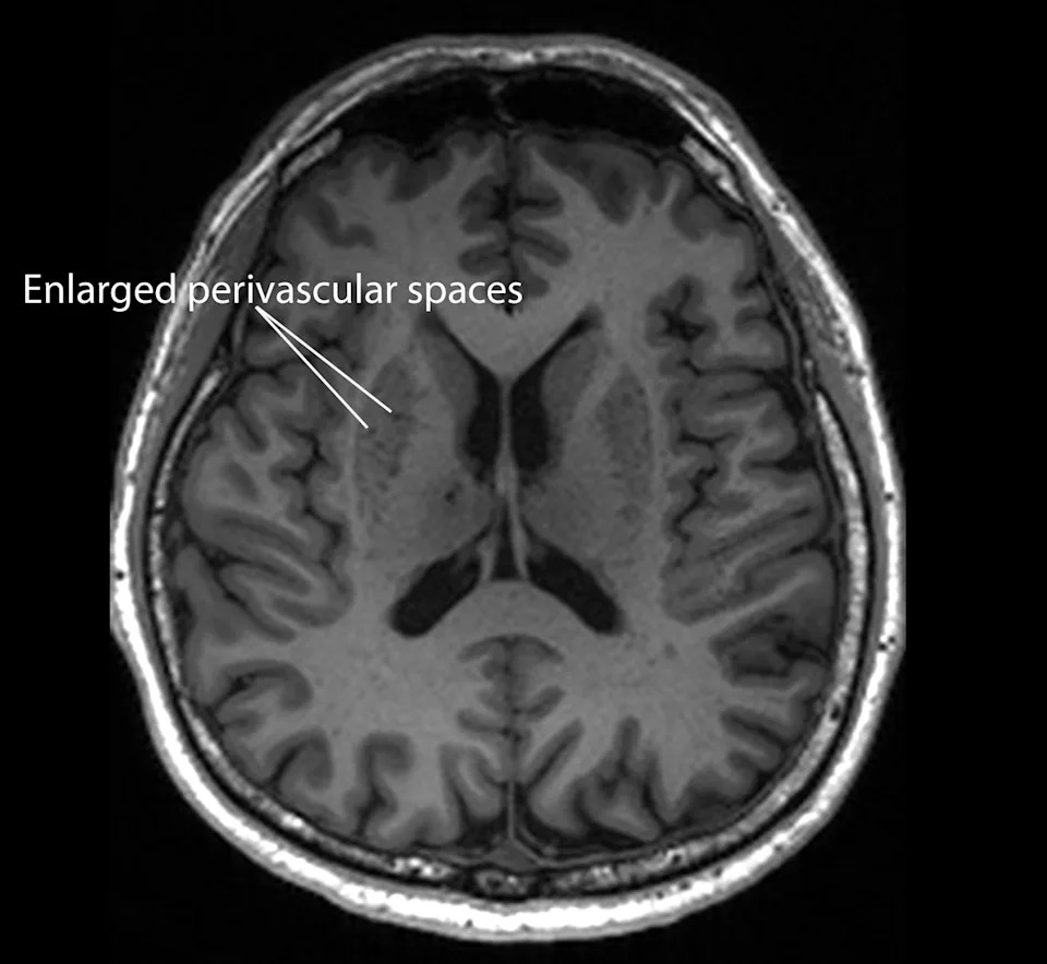
Magnetic resonance imaging image of a patient who has enlarged perivascular spaces, which are seen as dark lesions in dark grey regions around the center of the brain (NTU LKCMedicine)
Why This Matters
Alzheimer’s dementia progressively impairs memory, attention and daily functioning. With global dementia cases projected to rise substantially over the coming decades, low-cost, widely available screening tools could improve early detection and enable earlier interventions.
“These brain anomalies can be visually identified on routine magnetic resonance imaging (MRI) scans performed to evaluate cognitive decline,” said neurologist Nagaendran Kandiah of Nanyang Technological University, Singapore.
Limitations And Next Steps
Although the findings are promising, enlarged PVS on MRI are not a definitive diagnosis of Alzheimer’s. The study is observational and shows association rather than causation. Researchers plan prospective follow-up of the same participants to determine how many progress to Alzheimer’s and whether enlarged PVS reliably predict dementia over time.
Experts caution that further validation across diverse populations and standardized imaging criteria are needed before enlarged PVS can be used in routine clinical risk assessment. If confirmed, however, incorporating PVS assessment into routine MRI interpretation could be a cost-effective adjunct to blood biomarkers and other diagnostic tools.
Bottom Line
Enlarged perivascular spaces on routine MRI scans may represent an accessible early indicator of Alzheimer’s risk, but more research is required to confirm their predictive value and establish clinical guidelines for their use.
Help us improve.