Scientists built ChromAgeNet, an AI that classifies young versus old hematopoietic stem cells by analyzing 3D chromatin organization inside cell nuclei. Trained on microscope images from mice and reported in Aging Cell, the model detects subtle structural changes — like chromatin compactness and alterations at the nuclear periphery — that typical chemical age clocks miss. The approach is interpretable, letting researchers see which features drive classifications. While some epigenetic drugs made aged cells' chromatin look younger, the team cautions this does not prove functional rejuvenation and human validation is still needed.
AI Detects Hidden Aging Signature in Blood Stem Cells by Reading 3D DNA Architecture

Photo by Getty Images
Researchers have developed an artificial-intelligence tool that detects a previously hidden sign of cellular aging — not in chemical marks on DNA, but in the way DNA is folded and arranged inside the nuclei of blood-forming stem cells.
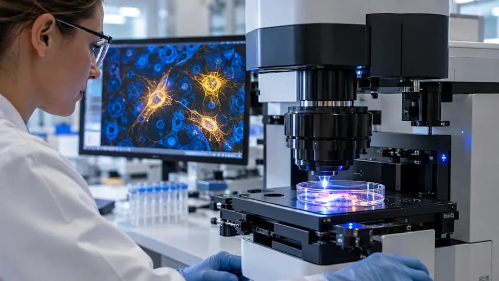
ChromAgeNet, described in the journal Aging Cell, analyzes three-dimensional microscope images of hematopoietic (blood-forming) stem cells from mice to distinguish younger from older cells. Unlike most biological-age clocks that rely on chemical changes such as DNA methylation, this model evaluates nuclear architecture — the physical organization and compactness of chromatin.
How the AI Works
The team trained ChromAgeNet on 3D images of chromatin organization. The algorithm learned to recognize subtle structural features that are often invisible to the human eye, including variations in chromatin compactness and alterations concentrated near the nuclear periphery. Importantly, the model is interpretable: researchers can inspect which image features most influenced each age classification, helping link visual patterns to biological hypotheses rather than relying on a black-box prediction.
"The DNA has a precise three-dimensional structure that changes over time," researcher Paula Petrone said in comments reported by EFE. The AI can pick up on age-related features "that are not necessarily perceived by the human eye."
Testing Interventions — Cautious Results
Beyond classifying age, the team used ChromAgeNet to examine whether interventions change nuclear architecture. When aged stem cells were treated with epigenetic drugs, some cells developed chromatin patterns more characteristic of younger cells. The authors emphasize a critical caveat: changes in chromatin appearance do not prove these cells regained youthful function. Functional assays are still required to determine whether structural changes translate into improved stem cell performance.
The experiments were performed in mice rather than humans. Maria Carolina Florian of the IDIBELL team told EFE that the researchers hope the method could one day help screen compounds that improve immune function and, ultimately, "the quality of life of older people."
Why This Matters
This work highlights a complementary direction for aging research: imaging-based assessment of nuclear architecture adds a structural dimension to chemical biomarkers. If validated in human cells and linked to functional outcomes, imaging tools like ChromAgeNet could help researchers better understand the mechanics of cellular aging and accelerate discovery of interventions that restore stem cell health.
Next steps include validating the approach in human samples, linking imaging signatures to functional measures of stem cell health, and testing whether drugs that alter chromatin structure can reliably rejuvenate cellular function.
Help us improve.