The Vrije Universiteit Amsterdam team created a three-layer human cell "tri-culture" placental model and found most tested micro- and nanoplastics crossed the membrane within 72 hours. Fluorescence imaging showed particles lodged mainly in the outer two cell layers, and the model produced 19 steroid hormones before exposure. After exposure, several polymers reduced etiocholanolone and polystyrene altered estrogen levels. The authors stress these results do not prove harm in pregnancies but call for urgent further study on mechanisms and developmental impacts.
Study: Micro- and Nanoplastics Cross a Human Placental Model and Alter Steroid Hormones
(Henrik5000/Getty Images)
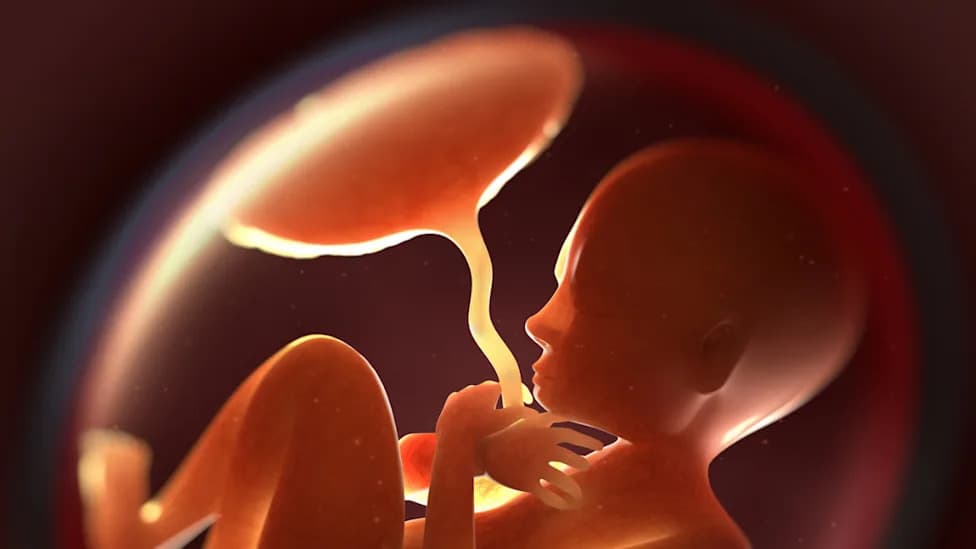
Tiny fragments of plastic are being found in more human tissues than previously thought, and a new laboratory study suggests several types of micro- and nanoplastics can cross a model of the human placental barrier and change steroid hormone production.
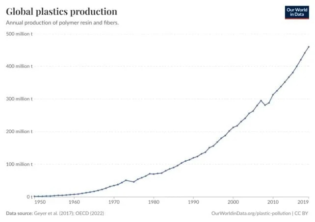
Global plastics production has been increasing since the 1950s. (Our World in Data)
How the Study Was Done
Researchers at Vrije Universiteit Amsterdam built a three-layer in vitro "tri-culture" placental model to better mimic key features of the human maternal–fetal interface. The outer layer used BeWo b30 cells (trophoblast-like epithelial cells), the middle layer used HUVEC cells (human umbilical vein endothelial cells), and the inner layer used H295R cells (human adrenal-derived cells representing the fetal adrenal steroid-producing compartment).

Subscribe to ScienceAlert's free fact-checked newsletter
Exposure and Measurement
The team applied several common plastic polymers (including polystyrene, PMMA, PVC, PET and polyamide) to the outer layer and tracked particle translocation and hormone output over 72 hours. Fluorescence microscopy assessed particle localization, and steroid hormone profiling measured production by the tri-culture system.
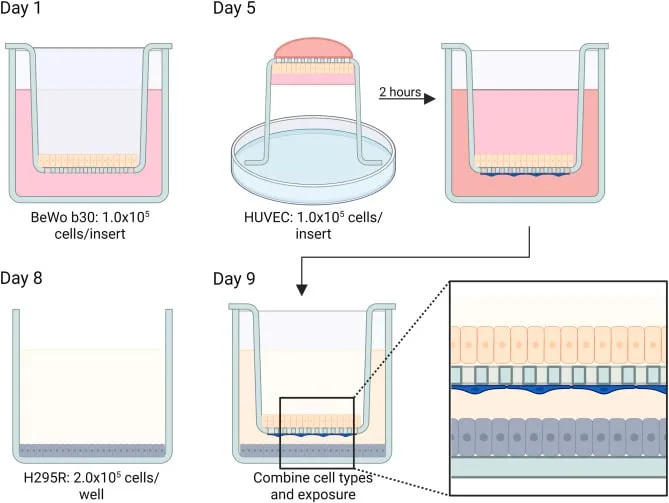
Schematic overview of the experimental procedure for establishing the tri-culture placenta model. (van Boxel et al.,Mol. Cell. Endocrinol., 2026)
Key Findings
- Most tested polymers traversed the tri-culture membrane to some degree within 72 hours; polyamide was the one polymer not detected in measurable amounts on the basolateral side.
- Fluorescence imaging showed particles concentrated in the first two cell layers (BeWo b30 and HUVEC) but not inside the H295R cells.
- Before exposure the model produced 19 distinct steroid hormones, indicating active steroidogenic pathways consistent with key aspects of human placental hormone biology.
- After polymer exposure several plastics were associated with reduced etiocholanolone levels, and polystyrene exposure was linked to altered estrogen levels.
Context and Limitations
Previous studies have detected micro- and nanoplastics in human blood, placentas, amniotic fluid and meconium. Animal studies have shown plastics can reach fetal tissues in rodents, but species differences in placental anatomy and steroidogenesis limit direct translation to humans. The tri-culture model improves relevance to human biology but does not fully reproduce the full complexity of an in vivo human pregnancy.
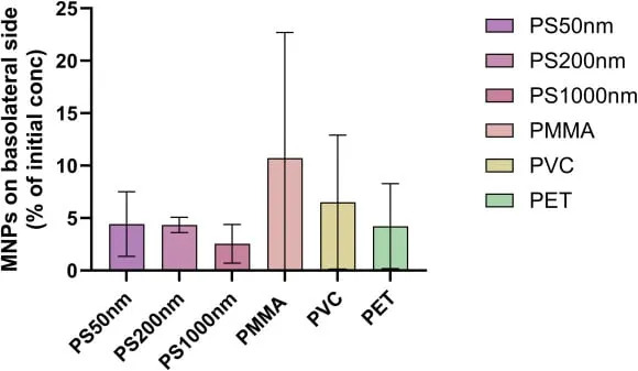
The bar chart shows the percentage of the initial concentration of each polymer applied to the placenta model that passed through to the basolateral layer, in comparison with vehicle-control-treated cells. PS = polystyrene; PMMA = poly(methyl methacrylate); PVC = polyvinyl chloride; PET = polyethylene terephthalate. (van Boxel et al.,Mol. Cell. Endocrinol., 2026)
"Our study shows that several types of micro- and nanoplastics can cross a human placental model and influence hormone production," said Jeske van Boxel, the study lead. "While this does not directly prove harm during pregnancy, it underscores the urgent need for further research on mechanisms and potential developmental consequences."
What This Means
The findings add experimental evidence that micro- and nanoplastics can cross a human-relevant placental barrier model and change steroid hormone profiles that play critical roles in fetal development. However, causation of adverse outcomes in pregnancies is not established and requires follow-up work using additional models, dose-response studies, and epidemiological investigation.
Publication: Molecular and Cellular Endocrinology (2026). Research team led by Jeske van Boxel, Vrije Universiteit Amsterdam.
Help us improve.