Researchers engineered fluorescent nanobodies that remain stable and functional inside living cells, enabling direct, multicolor visualization of intracellular proteins and processes. By embedding fluorophores into flexible internal loops of the nanobody, the probes preserve both binding and fluorescence and are available in blue, green, orange, red and near-infrared. The platform works in cultured human cells, neurons, mice and zebrafish embryos and supports simultaneous tracking of multiple targets. Delivering probes in vivo remains a challenge, but the advance transforms how scientists study cells alive.
Fluorescent Nanobody Toolkit Lets Scientists Watch Inside Living Cells in Multiple Colors
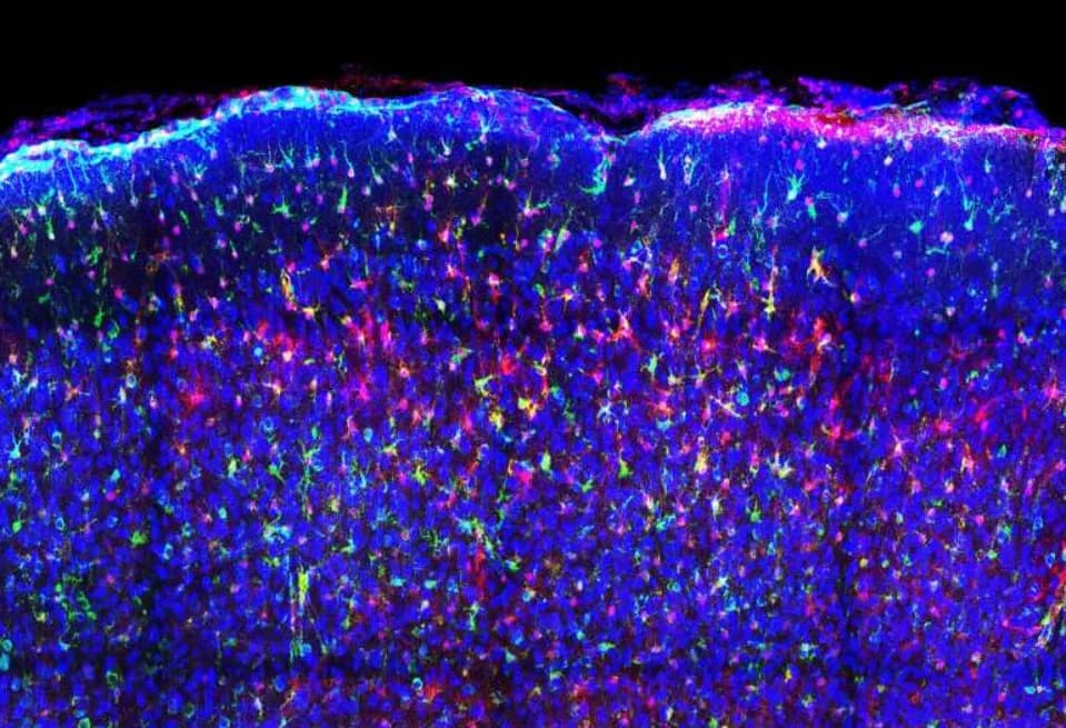
Multicolor image of mouse brain tissue showing astrocytes highlighted in purple and neurons shown in blue, illustrating cell-type-specific labeling.Credit: Nature Methods (2026). DOI: 10.1038/s41592-026-03056-3
Researchers have developed a new generation of fluorescent nanobodies—tiny, engineered antibody fragments—that can enter living cells, remain stable in the cytoplasm and light up specific intracellular targets. This advance gives scientists a practical way to observe molecular events inside intact cells, tissues and whole organisms without having to break cells apart.
How the Technology Works
Traditional antibodies are optimized to work in blood and extracellular fluids and typically fail inside cells: they misfold, aggregate or lose function. The new approach uses nanobodies, which are much smaller and more robust. Crucially, researchers embedded fluorescent labels into flexible internal loops of the nanobody rather than attaching them to the protein termini. That design preserves both binding activity and fluorescence, producing bright, stable probes that function inside living cells.
Color Palette and Specificity
The team created a palette spanning blue, green, orange, red and near-infrared probes. Each nanobody is engineered to recognize a specific intracellular target, from common lab marker proteins to viral or structural proteins. The probes are largely dark until they bind their targets, which sharply reduces background fluorescence and improves imaging specificity.
Demonstrated Applications
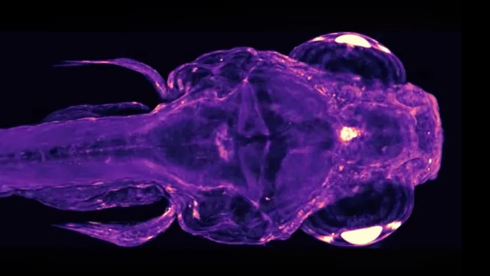
Tests in human cultured cells showed clear labeling of structures in the nucleus, at the cell membrane and within energy-producing compartments such as mitochondria. In neuronal preparations, the probes captured dynamic signaling events, including calcium fluctuations that underlie neuronal communication. The probes also worked in living animals: in mice they labeled distinct populations of brain cells and remained bright enough for deep-brain imaging, and in zebrafish embryos they reported activity in a key developmental signaling network.
Multiplexing and Biological Impact
Most intracellular imaging tools are limited to one or two colors. This platform supports simultaneous, multicolor imaging of several structures and target proteins within the same cell, enabling researchers to map interacting processes in three dimensions and over time. Because diseases typically involve multiple disrupted pathways, the ability to watch several processes at once offers a more complete view of disease mechanisms.
Limits and Future Directions
A major remaining challenge is efficient and safe delivery of fluorescent nanobodies into cells in vivo for clinical use. Despite that hurdle, the immediate impact for basic research is substantial: scientists can now observe intracellular processes in living cells and organisms without cell lysis or destructive preparation steps. Continued work will focus on delivery methods, improving probe brightness and expanding the library of target-specific nanobodies.
Context: This report is the third installment in a series on nanobody applications in medicine. Originally published on Forbes.com.
Help us improve.