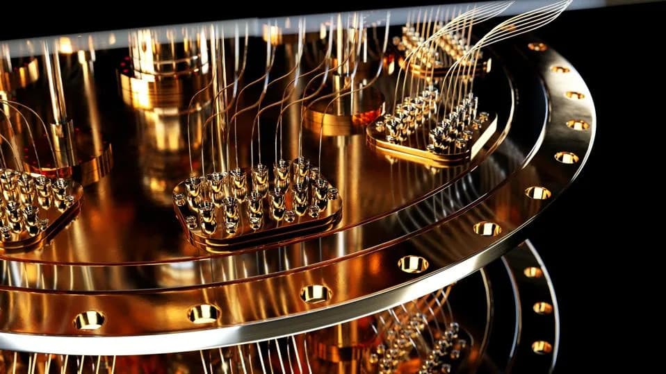
Gold nanostars positioned near membrane‑embedded QuasAr proteins increased fluorescence and unexpectedly sped up voltage reporting in living HEK293T cells. Coupling raised brightness (up to ≈2.4× for QuasAr1 and ≈1.69× for QuasAr6a in optimal placement) and accelerated photocycle kinetics (upward rates ≈230 s⁻¹ → ≈1,300 s⁻¹), while relative voltage sensitivity dropped from 0.37 to 0.11. The effect stems from plasmonic enhancement of absorption and emission, but precise nanoparticle placement and translation to neurons remain challenges.
Gold Nanostars Boost Brightness and Speed of QuasAr Voltage Sensors in Living Cells

Plasmonic nano-antennas altered a protein's light emission and response speed without changing its genetic code. (CREDIT: Shutterstock)
Gold nanostars placed near engineered voltage‑sensing proteins made those proteins both brighter and unexpectedly faster at reporting membrane voltage in living mammalian cells—without changing the proteins’ genes. Researchers at Delft University of Technology report in Advanced Materials that plasmonic coupling between colloidally grown gold nanostars and membrane‑embedded fluorescent proteins altered both emission intensity and photocycle kinetics, offering a physical route to improve genetically encoded voltage indicators.
What the Team Did
The investigators targeted Archaerhodopsin‑derived proteins, focusing on QuasAr6a (and also testing QuasAr1), expressed in HEK293T cells. They designed gold nanostars as plasmonic nano‑antennas tuned near the proteins’ excitation/emission range, and validated the design first with the dye Cyanine‑5 (Cy5) and in artificial lipid membranes before moving into living cells.
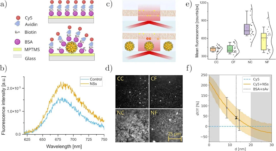
Control over nanostar-fluorophore coupling allows tunable plasmonic enhancement. (CREDIT: Advanced Materials)
Key Experimental Observations
Fluorescence enhancement (bench tests): Direct coupling of Cy5 to nanostars increased fluorescence by ~1.49× versus control; nanostars in artificial lipid membranes gave ~1.9×. Increasing separation (adding fibronectin, from ≈8.5 nm to ≈14.5 nm) reduced enhancement to ~1.3×.
Fluorescence in living cells: As nanostars accumulated near HEK293T cells, QuasAr1 brightness rose to ≈2.4× baseline and QuasAr6a rose generally to ≈1.3×. Placement mattered: nanostars immobilized beneath cells (with fibronectin) increased QuasAr6a to ≈1.69×, while nanostars deposited above cells produced ≈1.28×. Estimated separations were ≈13.5 nm (beneath) vs ≈16 nm (above).
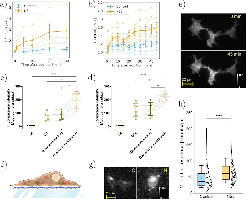
Gold nanostars enable plasmonic enhancement of QuasAr fluorescence in living cells. (CREDIT: Advanced Materials)
Kinetics and sensitivity: Voltage‑clamp steps from −70 mV to +30 mV showed accelerated QuasAr6a kinetics when coupled to resonant nanostars: upward response rate increased from ≈230 s⁻¹ to ≈1,300 s⁻¹ and the downward rate from ≈270 s⁻¹ to ≈930 s⁻¹. However, relative voltage sensitivity dropped from 0.37 to 0.11 (≈71% reduction), mainly because the baseline fluorescence increased.
Controls and safety checks: Only nanostars with the correct optical resonance produced the combined brightness/kinetic effects; 50‑nm nanostars and 90‑nm gold nanospheres did not. The team measured no significant additional heating under the illumination used.
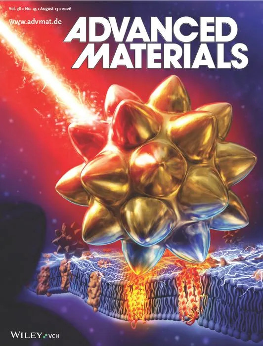
Cover illustration showing a gold nanostar illuminated by laser light. (CREDIT: 2026 Wiley-VCH GmbH)
How Plasmonic Coupling Explains the Effects
To explain the faster responses the authors built a four‑state model of the QuasAr6a photocycle. The best fit required two concurrent effects: an increase in fluorescence emission and an accelerated light‑driven transition. In the model that transition rate rose from ≈443 s⁻¹ (no nanostars) to ≈1,184 s⁻¹ (with nanostars), with an inferred field enhancement factor of ≈1.34. The nanostars’ broad resonance peaked near ≈740 nm, overlapping the 639‑nm excitation and the ~660–800 nm emission band—consistent with simultaneous enhancement of absorption and emission.
Limitations and Next Steps
Important uncertainties remain for translation to neurons or in vivo settings. The exact nanoscale positions of individual nanostars relative to proteins were not determined: some particles may have entered cells, and others likely acquired protein coronas in culture medium. Reported electrophysiological rates are averaged across cells rather than measured for individual nanoscale protein–particle pairs. Those factors matter because brightness, response speed and voltage sensitivity all influence whether a fluorescent sensor can resolve brief electrical events.
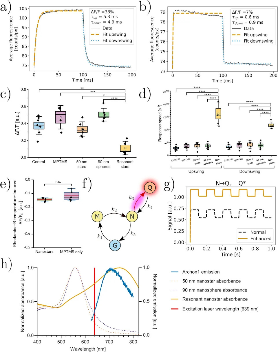
Plasmonic enhancement by gold nanostars manipulates the photocycle of QuasAr6a and modifies its response to voltage. (CREDIT: Advanced Materials)
Future work will need to refine nanoparticle resonance, spacing and placement and to test the approach in neuronal preparations and brain tissue. The study demonstrates a complementary, physical engineering route—tuning the optical environment around an evolved protein—that can produce concurrent gains in brightness and temporal resolution, albeit with trade‑offs in sensitivity.
Bottom line: Nearby resonant nanostructures can alter a functional protein’s photophysics inside living mammalian cells, accelerating photocycle steps and enhancing emission—but practical application will require precise control of spacing, resonance and placement.
References: Research published in Advanced Materials; experiments performed in HEK293T cells; modeling and control experiments described in the paper.
Help us improve.