Researchers transformed a DCM patient’s blood cells into iPSCs and then cardiomyocytes, assembling them into a 3D, beating heart-on-a-chip that reproduced structural and functional hallmarks of dilated cardiomyopathy, including irregular beating. Fluorescent beads mapped local contractility and the tissue responded to norepinephrine, showing physiological responsiveness. The study used one patient and one healthy control, so findings are preliminary and require validation in larger, diverse cohorts before clinical use.
Patient's Blood Cells Grow Beating 'Mini-Heart' That Mirrors Dilated Cardiomyopathy
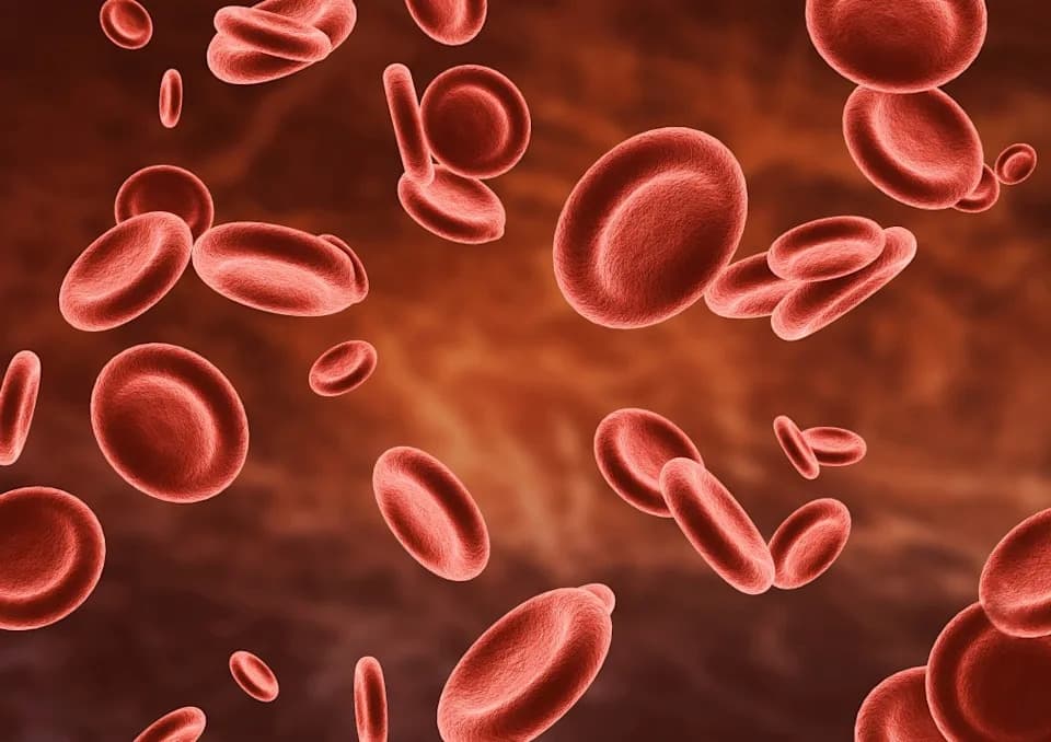
A stock image shows a group of human red blood cells in the bloodstream microscopic view.
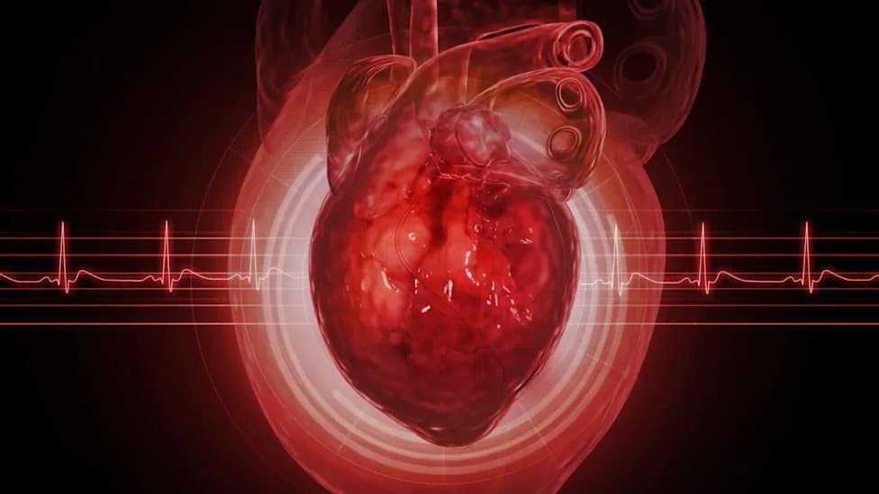
Scientists have engineered a tiny, beating piece of heart tissue from a patient’s own blood cells, producing a proof-of-concept model that reproduces key features of dilated cardiomyopathy (DCM). The lab-grown, patient-specific “mini-heart” contracted on its own and showed structural, electrical and molecular differences consistent with the disease.
How the Mini-Heart Was Made
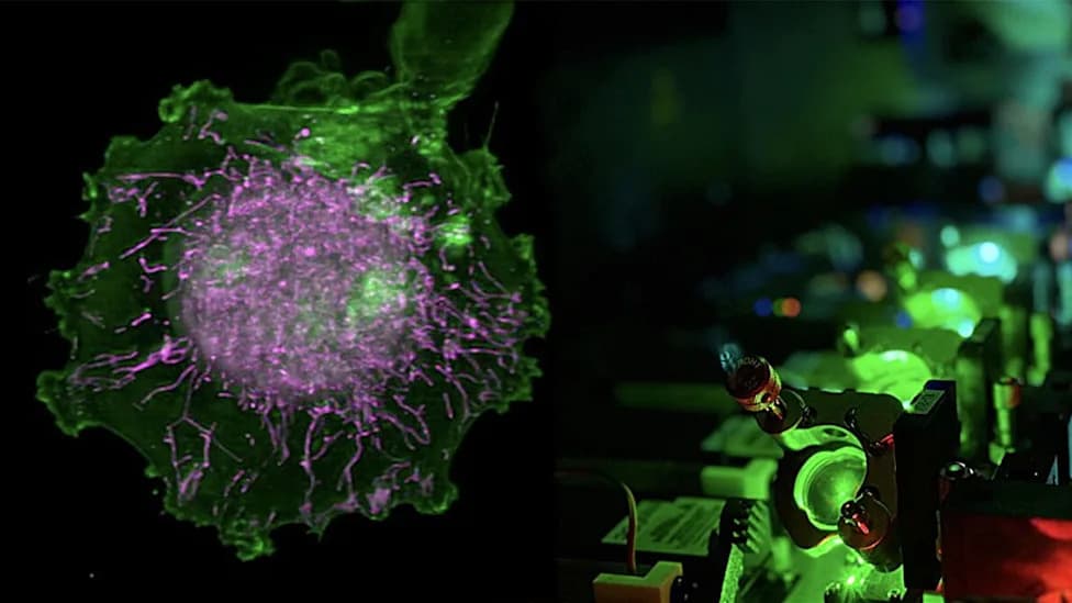
Researchers collected blood from an individual diagnosed with DCM and reprogrammed those blood cells into induced pluripotent stem cells (iPSCs). The iPSCs were then differentiated into cardiomyocytes — the specialised muscle cells that drive the heart’s contractions — and assembled into a three-dimensional heart tissue on a chip. Tiny fluorescent beads were embedded throughout the tissue to map local movement and measure contractility across regions during each beat.
Direct Comparison With Healthy Tissue
To test disease relevance, the team built a second heart-on-a-chip using cells from a healthy donor and compared the two models side by side. The DCM-derived tissue showed abnormal and irregular beating, altered calcium dynamics, distinct contraction patterns and molecular signatures that differed from the healthy control. Both tissues responded to norepinephrine stimulation, demonstrating that the engineered models react to physiological cues.
Dr. Ayesha Bryant, Clinical Advisor at Alpas Wellness, told Newsweek: “These patient-derived models may reveal insights unavailable from animal models or simple cell cultures because they reflect an individual’s unique genetic makeup. While promising, they are not yet ready to guide clinical treatment choices.”
Implications and Limitations
The platform could accelerate mechanistic studies and drug development for DCM, help prioritise candidate therapies earlier in the pipeline and potentially reduce reliance on some animal studies. Importantly, this study is an early demonstration: it used cells from a single DCM patient and one healthy control, so the findings are preliminary. Broader validation across many patients with diverse genetic backgrounds and DCM subtypes will be essential before clinical application or personalized-treatment predictions are possible.
Reference: Ali Mousavi et al., Patient-Derived 3D Heart-On-a-Chip Model of Dilated Cardiomyopathy With Embedded Bead-Based Mapping of Tissue Contractility, Advanced Healthcare Materials (2026). DOI: 10.1002/adhm.71464.
Help us improve.