Researchers at UC San Diego combined 4D lattice light-sheet microscopy, deep learning and physics-based digital twins to study mitochondrial dynamics. They built MitoSpace from roughly 40,000 single-cell movies of cancer cells treated with 25 mitochondrial-disrupting compounds, enabling the system to cluster drug responses without manual labels. A complementary rules-based digital twin reproduced mitochondrial reactions seen in living cells, suggesting some early screening could move in silico. The approach aims to speed and add biological depth to early-stage drug evaluation for diseases linked to mitochondrial dysfunction.
AI and 40,000 Live-Cell Movies: Digital ‘Cell Twins’ Reveal How Drugs Reshape Mitochondria
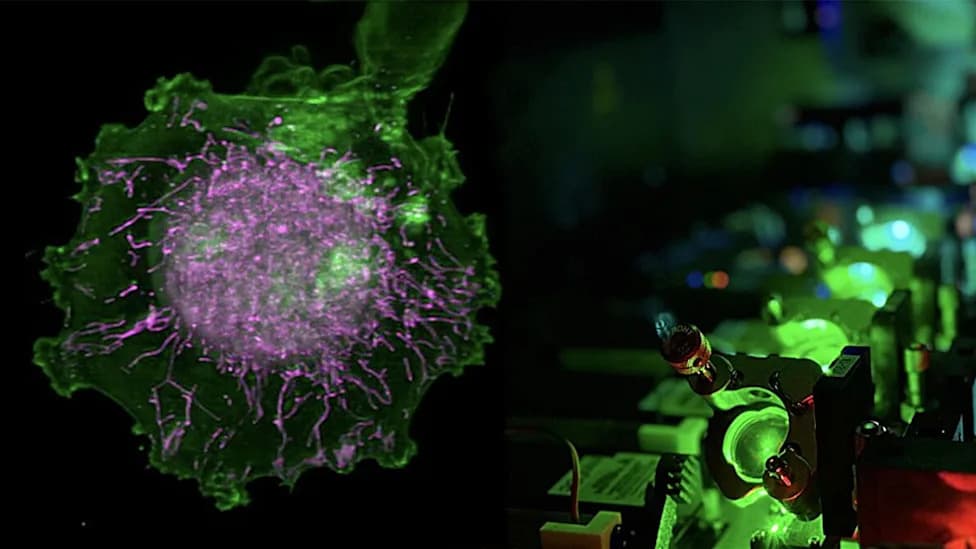
Photo Credit: UC San Diego Health Sciences
Mitochondria are often depicted as static, bean-shaped blobs in textbooks, but inside living cells they form a dynamic, constantly remodeling network. Researchers at the University of California San Diego combined advanced 4D microscopy, deep learning and physics-based “digital twin” simulations to study how drugs alter mitochondrial form and function at unprecedented scale.
What the team did
The researchers used 4D lattice light-sheet microscopy to record roughly 40,000 single-cell movies that capture mitochondrial and organelle motion through three-dimensional space over time. They exposed cancer cells to 25 compounds known to perturb mitochondria by distinct mechanisms and assembled those time-resolved movies into a dataset named MitoSpace.
From this dataset they developed two complementary tools:
- MitoSpace (deep learning) — a neural network trained to interpret mitochondrial morphology and dynamics as a proxy for cell health. The model autonomously learned recurring patterns and could cluster cells by drug response even without labels identifying each treatment.
- Rules-based digital twin (physics model) — a simulation that models organelle behavior and mitochondrial network dynamics inside a virtual cell. When drugs were applied in silico, the simulated networks reacted in ways that paralleled changes observed in living cells.
Why this matters
Because mitochondrial shape and dynamics carry signals about cellular state — healthy, stressed, or damaged — capturing their motion provides richer information than single-frame, two-dimensional images. Turning moving organelles into computational models allows researchers to screen and characterize promising compounds more rapidly in silico before committing to slower, costlier bench experiments.
Limitations and next steps
The work is early-stage and intended as a research tool rather than a finished therapy or consumer product. The models must be validated across more cell types, disease contexts, and compound classes. If the approaches prove robust, they could reduce labor and accelerate early-stage screening, particularly for diseases where mitochondrial dysfunction is a component (for example, cancer, diabetes, Alzheimer’s disease and pediatric mitochondrial disorders).
Context and related work
These studies—reported in the journal Cell—join broader efforts applying AI to biology, including virtual-animal models, protein language models for drug discovery, and AI forecasting of flu strains. The authors emphasize these methods are intended to help scientists prioritize and interpret candidates with richer biological context than static imaging alone.
Bottom line: Combining 4D imaging, deep learning and physics-based digital twins provides a scalable way to read mitochondrial dynamics as a window into cell health and drug response, offering a promising route to faster, more informative early-stage drug evaluation.
Help us improve.