Researchers show that adults — both blind and sighted — can learn click-based echolocation after roughly 10 weeks of training, using mouth clicks or cane taps. Brain scans of 26 trainees revealed functional and structural changes in primary sensory areas: V1 (visual cortex) became sensitive to sound echoes while A1 also adapted. Comparative cetacean research finds dolphins have stronger cerebellar connections, underscoring that echolocation-related adaptations can appear in unexpected brain regions. Continued cross-species and longitudinal studies will deepen our understanding of multisensory plasticity.
Humans Can Learn Click-Based Echolocation in about 10 Weeks — Training Rewires the Brain
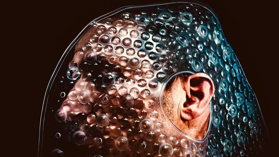
(Sebastian Schuster/Unsplash)
Echolocation — producing sounds and interpreting returning echoes to map the environment — is best known in bats and dolphins. Recent human studies show that adults, both blind and sighted, can acquire click-based echolocation with relatively short training, and that learning produces measurable changes in the brain.
A 2021 study published in PLOS One by researchers at Durham University trained volunteers to use mouth clicks (and, in everyday practice, some people use cane taps) to detect objects and spatial layouts. After approximately 10 weeks of systematic training, many participants could reliably use echoes to orient themselves and identify nearby obstacles.
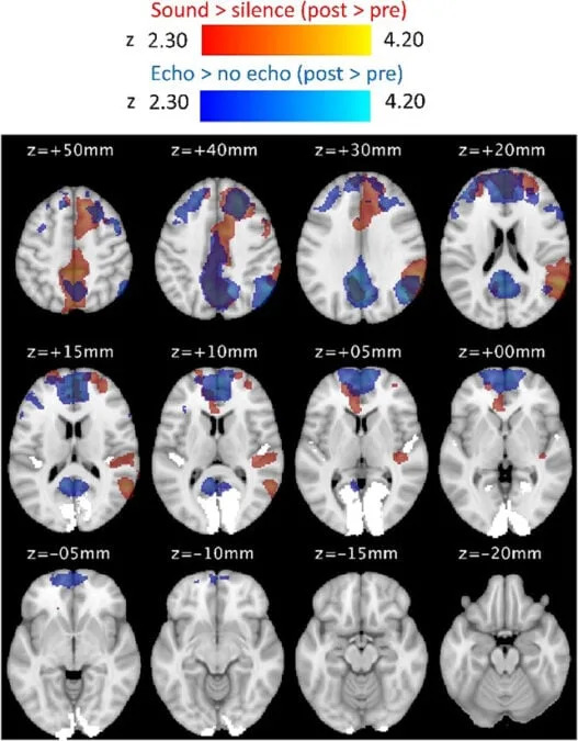
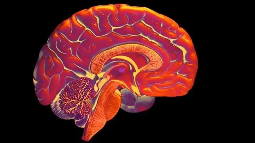
Researchers wanted to see how echolocation training changed the brain – and the differences were similar across blind and sighted participants. (Norman et al.,Cereb. Cortex, 2024)
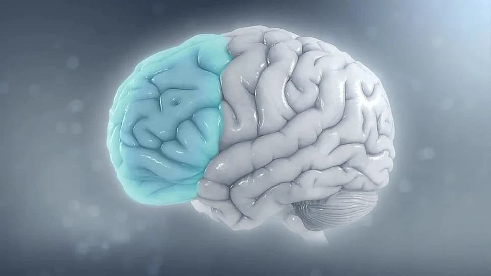
A follow-up study published in Cerebral Cortex examined how that same 10-week training altered brain structure and function in the 26 participants who completed the program. The team focused on two primary sensory areas: the primary visual cortex (V1), normally associated with vision, and the primary auditory cortex (A1), associated with sound processing.
Strikingly, functional and structural imaging revealed that V1 developed sensitivity to sound echoes in both blind and sighted trainees. In other words, a brain region typically dedicated to visual input began responding to auditory echo information after training.
YouTube Thumbnail
"We show here, for the first time, functional and structural brain changes in primary sensory areas V1 and A1 in blind and sighted people who learn click-based echolocation in adulthood," the authors write.
The overall pattern of cross-modal plasticity was similar across blind and sighted participants, indicating that long-term sensory deprivation is not required for a primary sensory area to adopt sensitivity to a new modality. The investigators do note, however, that some specific structural changes differed between blind and sighted trainees, suggesting sensory history still shapes the exact rewiring.
Comparative Insights From Cetaceans
Complementing the human work, a 2025 PLOS One comparative study examined how evolution shaped echolocation-related circuitry in cetaceans. Researchers compared brains from three dolphins (echolocating species) and one baleen whale (a non-echolocating species that still relies heavily on sound).

Subscribe to ScienceAlert's free fact-checked newsletter
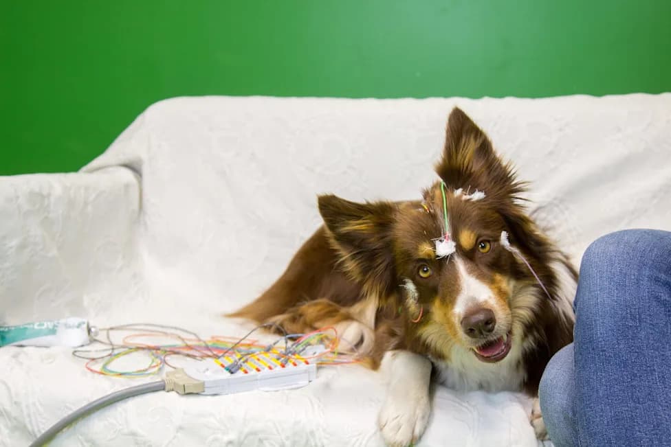
While many auditory features were similar across species, dolphins showed notably stronger connections from auditory regions to the cerebellum. As biologist Peter Tyack explains, the cerebellum is increasingly recognized as a center for rapid prediction and integration of sensory and motor information — functions that would be useful for precise, fast echolocation.
Together, the human and cetacean findings highlight that adaptations for echolocation can appear in brain regions not traditionally associated solely with hearing or vision, and that both learning and evolution can reshape neural circuits to combine senses in new ways.
The studies discussed here were published in PLOS One and Cerebral Cortex. Further research across larger groups, different training methods, and additional species will help clarify how echolocation skills are acquired and how multisensory brain networks reorganize in response.
Help us improve.