
The zap-and-freeze method freezes brain tissue milliseconds after electrical stimulation, enabling visualization of ultrafast synaptic events. Johns Hopkins researchers observed endocytosis occurring in under 100 milliseconds in both mouse and human brain slices and identified dynamin1xA as critical to this rapid recycling. The team plans to study tissue from Parkinson's patients to look for disease-related synaptic differences. Results are published in the journal Neuron.
Zap-and-Freeze Captures Millisecond Synaptic Events — Could Shed Light On Parkinson's

Biology nerve cell with biomedicine concept
Researchers have refined a high-speed imaging technique that freezes brain tissue within milliseconds of electrical stimulation, revealing ultrafast synaptic events previously too rapid to study. The method, called zap-and-freeze, couples a brief electrical jolt with high-pressure freezing to lock neurons and their membrane dynamics at precise moments of activity.
How Zap-and-Freeze Works
Led by teams at the Johns Hopkins University School of Medicine, investigators applied zap-and-freeze to brain slices from both mice and humans. The high-pressure freezing preserves tissue structure and function while capturing snapshots of active synapses for detailed electron-microscopy analysis.
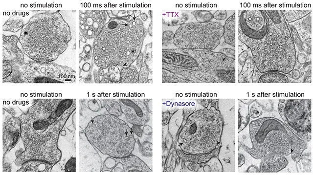
The researchers imaged cell activity milliseconds after stimulation. (Eddings et al.,Neuron, 2025)
Key Findings
The experiments revealed direct evidence of ultrafast endocytosis — the rapid recycling of vesicle membrane after neurotransmitter release — occurring in under 100 milliseconds in both mouse and human samples. The team also identified the protein dynamin1xA as essential to that rapid recycling process.
"This approach has the potential to reveal dynamic, high-resolution information about synaptic membrane trafficking in intact human brain slices," said Johns Hopkins neuroscientist Chelsy Eddings and colleagues in their paper.
Implications For Parkinson's Disease
Because synaptic signaling and vesicle recycling are fundamental to neuronal communication, better resolving these processes could help researchers understand mechanisms that go awry in neurodegenerative disorders such as Parkinson's disease. While Parkinson's involves progressive neuron loss and complex pathology, comparing synaptic dynamics in healthy and affected tissue could clarify whether and how faulty vesicle trafficking contributes to disease progression.

Subscribe to ScienceAlert's free fact-checked newsletter
The researchers plan to obtain, with appropriate consent, tissue from patients undergoing invasive brain procedures for Parkinson's and other conditions to compare vesicle activity directly between healthy and diseased tissue.
Why It Matters
Zap-and-freeze joins a suite of modern techniques that allow neuroscientists to capture fleeting molecular events in near-living brain tissue. That temporal and structural resolution strengthens the link between animal models and human biology and may accelerate discovery of disease mechanisms and, eventually, therapeutic targets. The study is published in the journal Neuron.


