UC San Diego researchers propose that rhythmic muscle contractions in the gut — modeled as coupled oscillators — reveal how tiny brain arterioles synchronize their pulsatile dilation. Published in Physical Review Letters, the study shows how neighboring oscillators can lock frequencies and produce step-like transitions. This mathematical insight may advance understanding of cerebral blood flow regulation and gastrointestinal motility disorders.
Gut Muscle Rhythms May Explain How Tiny Brain Vessels Sync Their Pulses

Gut rhythms offer a clue to how brain blood vessels coordinate
The human body hosts many built-in rhythms — from the sleep–wake cycle to the steady pulsing of blood through the brain and the heartbeat. New research from the University of California San Diego suggests that rhythmic contractions in the gut may help explain how tiny blood vessels in the brain sync their pulses.
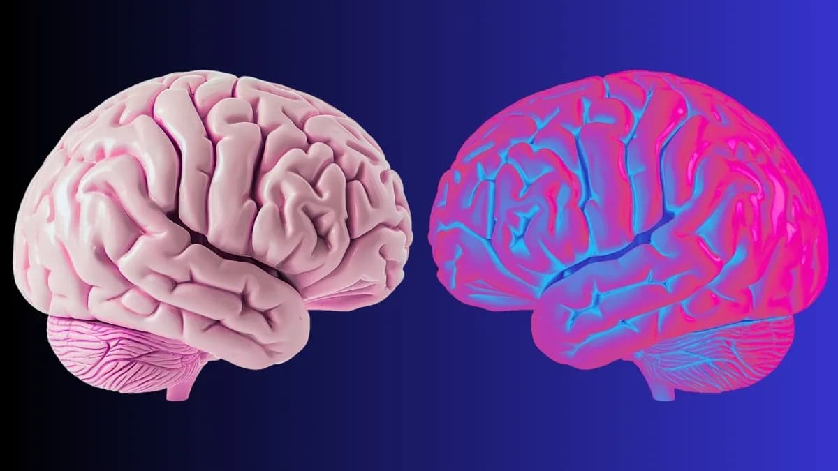
When neurons become active, small blood vessels called arterioles dilate to supply extra oxygen and nutrients. These arterioles often pulse in a coordinated way, but the mechanism that produces that coordination has been unclear.
The UC San Diego team drew an analogy with the digestive tract. The intestines move food by contracting in waves, and the researchers modeled intestinal segments as a chain of coupled oscillators that produce a staircase-like pattern of activity. Using mathematical analysis published in Physical Review Letters, they showed how neighboring oscillators can lock onto one another when their natural frequencies are similar, producing abrupt, step-like transitions between synchronized states.
“Coupled oscillators talk to each other and each section of the intestine is an oscillator that talks to the other sections near it,” said Massimo Vergassola, professor of physics at UC San Diego.
The same mathematical mechanism, the authors propose, could underlie how arterioles across nearby regions of the brain align their rhythms to deliver blood precisely where and when it is needed. David Kleinfeld, professor of physics and neurobiology at UC San Diego, emphasized that the new analysis resolves details of the transitions between locked states that previous approximations missed.
“The mathematics had been solved in an approximate way before now, but not in a way that gave you these breaks and what happens at the breaks. That’s a critical discovery,” Kleinfeld said.
Implications of this work extend beyond basic brain physiology. The findings could inform future studies of cerebral blood flow regulation and improve understanding of gastrointestinal motility disorders that disrupt the coordinated movement of food, liquids and waste through the gut.
For readers who want further background, Johns Hopkins Medicine provides accessible information on the brain–gut connection.
Help us improve.