Masakazu Iwasaka reports in a bioRxiv preprint that the skin of cobalt silversides (Hypoatherina tsurugae) shows rapid, reversible changes in reflected light that suggest active response rather than passive reflection. Iridophore cells filled with guanine crystals switch among "static bright," "dynamically twinkling," and "dark" states when illumination is applied or removed. The quenching response is strongest under blue light and commonly recovers within ~10 seconds, prompting hypotheses about neural modulation or opsin involvement; however, the study is preliminary and not yet peer-reviewed.
Fish Skin That 'Senses' Light: Cobalt Silversides Show Rapid, Blue-Sensitive Iridophore Responses
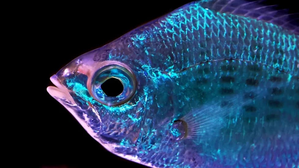
A tropical fish reflecting light. (David Clode/Unsplash)
A glittering school of silverside fish can look like a disco ball: countless tiny mirrors creating a dazzling shimmer. New laboratory observations reported in a bioRxiv preprint suggest that the skin of the cobalt silverside (Hypoatherina tsurugae) may do more than reflect light — it may actively respond to changes in illumination.
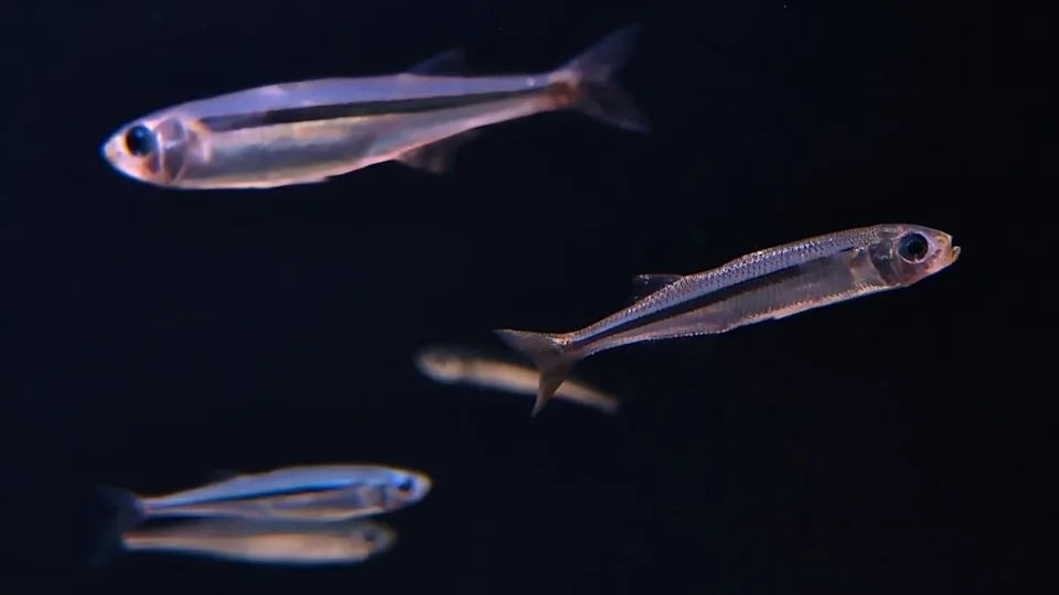
Hypoatherina tsurugae. (Nils Robert/iStock/Getty Images)
Key Observations
Masakazu Iwasaka, an interdisciplinary engineer at Hiroshima University, examined microscopic structures in the skin of 22 wild-caught cobalt silversides kept in a laboratory aquarium. These fish have dense clusters of iridophore cells packed with plate-like guanine crystals. Unlike pigment-based color in human skin, the crystalline stacks in iridophores produce color by bending and reflecting light at particular angles and wavelengths.
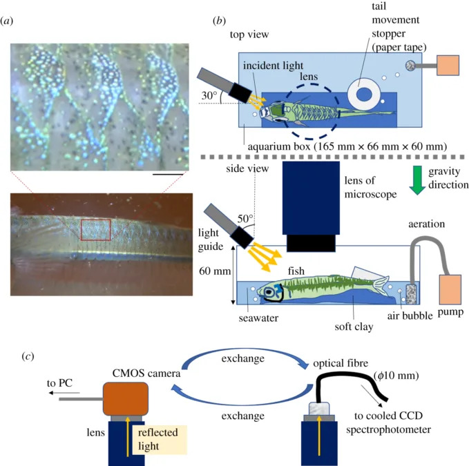
Methods used to inspect the fish's iridophores, as established in an earlier experiment. (Iwasaka,R. Soc. Open Sci., 2021)
In earlier work, Iwasaka documented spots of iridophores along the dorsal trunk that produced rapid, repetitive reflections at frequencies of several hertz independent of body motion. In the current experiments those same iridophore patches were observed to switch between three distinct visual states, which he described as "static bright," "dynamically twinkling," and "dark."
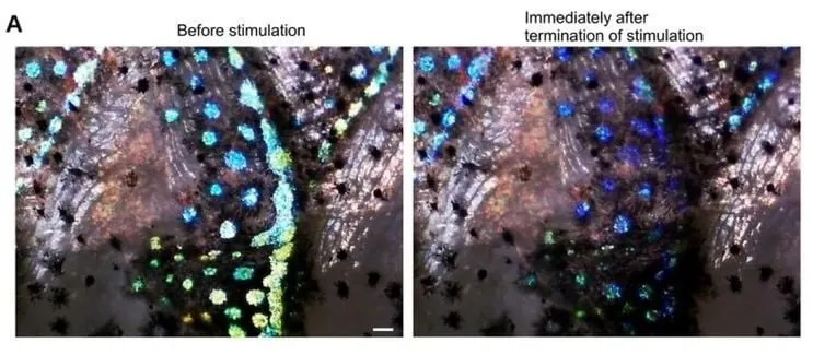
Quenching of light reflection from iridophores immediately after white LED stimulation. (Iwasaka,bioRxiv, 2026)
Controlled Light Tests
To test whether ambient light controls those states, some fish were briefly anesthetized and examined with a microscope-equipped camera while the investigator varied illumination. When a white LED (matching the aquarium/room lighting) was directed at targeted skin patches, the iridophores held the static bright appearance. When the direct light was switched off, those patches rapidly transitioned to the dark state — reflecting far less light — and then returned to bright or twinkling modes within seconds as ambient light readjusted.

Subscribe to ScienceAlert's free fact-checked newsletter
Iwasaka repeated the tests using blue, green and red LEDs, plus a blue laser and two types of green lasers. Spectral analysis showed the quenching (rapid drop in reflected light when illumination stopped) was most sensitive to blue wavelengths, weaker under green, and weakest under red. Across trials, iridophores typically recovered to a twinkling or active state about 10 seconds after the direct light source was removed.
Possible Mechanisms and Cautions
The speed and reversibility of these transitions suggest mechanisms faster than slow intracellular structural rearrangements. Iwasaka proposes that rapid neural modulation or intrinsic photoreceptive proteins such as opsins — which are best known from eyes and retinas — could contribute. However, the study did not test molecular or neural mechanisms directly.
Important: This work is reported in a preprint uploaded to bioRxiv and has not yet undergone peer review. The results are intriguing but provisional and should be interpreted cautiously until validated by further studies.
Why It Matters
If confirmed, these findings would expand our understanding of structural coloration in marine animals and suggest that some skin-based color systems can also function as light sensors — potentially informing behavior, camouflage, or communication. The observations raise new questions about the evolution, neural control, and molecular basis of light-responsive skin in fishes.
Help us improve.