Researchers identified ruptoblasts, a glandular cell type in planarian flatworms that defends tissue by rupturing in a process called ruptosis. Triggered by elevated activin (ACT-2), about 60–70% of ruptoblasts burst within roughly five minutes, releasing diffusible toxins that kill nearby cells and about 45% of adjacent bacteria. Ruptoblasts contribute to infection defense and a transplant-like tissue-rejection response; their gene signatures suggest an ancient bilaterian origin. Key molecular details remain unresolved.
Explosive 'Ruptoblast' Cells: A Fast, Sacrificial Immune Defense Found in Planarians
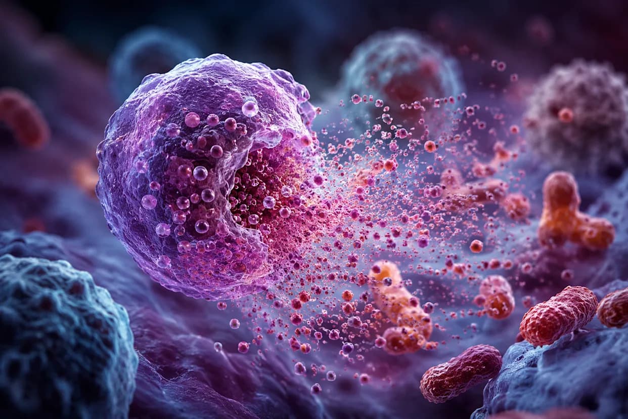
Flatworms use newly identified ruptoblasts, exploding cells triggered by activin, to kill bacteria and reject foreign tissue. (CREDIT: AI-generated image / The Brighter Side of News)
Researchers have discovered a previously unrecognized cell type in the planarian flatworm Schmidtea mediterranea that defends tissue by rupturing and releasing a potent chemical burst that kills nearby bacteria and damaged cells within minutes. The team named these gland-like cells "ruptoblasts" and the rapid, one-time death they undergo "ruptosis." Unlike conventional immune cells derived from white-blood-cell lineages, ruptoblasts represent a distinct, fast, sacrificial defense strategy.
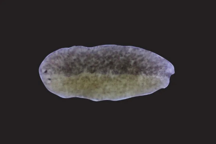
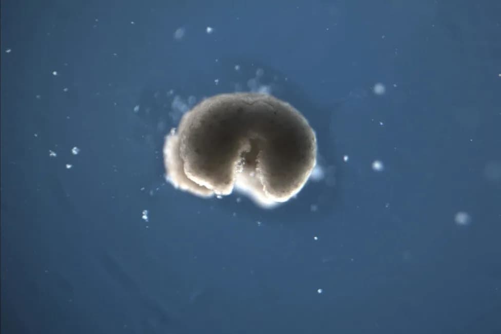
A chimera planarian worm fused from two different strains of planarians. (CREDIT: Wang Lab)
The study, published in the journal Cell, was led by Prof. Benyamin Rosental (Ben-Gurion University of the Negev) and Prof. Bo Wang (Stanford University). What makes this response striking is its trigger: a rise in the hormone activin—specifically a planarian form called ACT-2—normally involved in regeneration, reproduction, and tissue homeostasis. When activin spikes, ruptoblasts respond within minutes with an explosive lysis that the authors call ruptosis, producing a tightly confined zone of destruction.
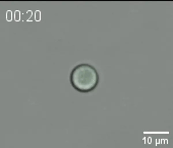
A video of ruptosis in action. (CREDIT: Wang Lab)
How the Team Discovered Ruptoblasts
The researchers first noted that injecting ACT-2 into planarians activated molecular pathways linked to inflammation and produced localized tissue damage and elevated cell death. To identify the effector cells, they dissociated animals into single cells and exposed the cells to ACT-2. A distinct granular population lysed within about five minutes; control proteins did not trigger this reaction. Follow-up single-cell RNA sequencing and targeted experiments tied the effect specifically to a glandular cell cluster: roughly 60–70% of those cells ruptured when exposed to activin, discharging granules that dispersed and rapidly vanished.
ACT-2 induces inflammatory responses in planarians. (CREDIT: Cell)
What Ruptoblasts Do
Ruptoblasts differ from familiar immune tactics. T cells and natural killer cells usually kill through direct contact; neutrophils can release extracellular DNA traps, a slower and mechanistically different response. Ruptoblasts do not make DNA traps and do not seem to require direct contact with targets. Instead, imaging showed nearby cells dying soon after a ruptoblast burst, consistent with release of diffusive toxic agents that act across a short radius.
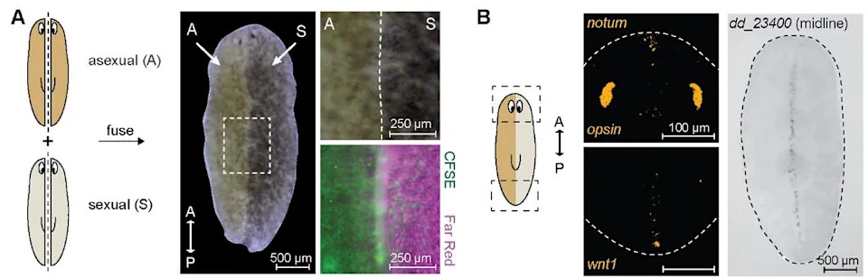
Genetic chimerism induces activin overactivation, resulting in chronic inflammation. (CREDIT: Cell)
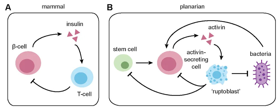
In live animals, ACT-2 injections wiped out ruptoblasts at the injection site and also eliminated neighboring activin-secreting cells and neoblasts (the stem cells that enable planarian regeneration) within roughly 100 micrometers. That combination suggests ruptoblasts can remove both problematic cells and the cells that might replenish them.
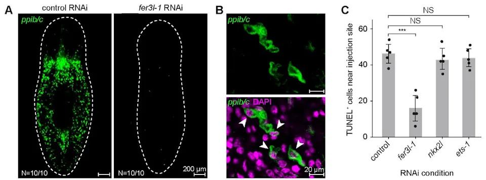
Ruptoblast-mediated cytotoxicity in vivo. (CREDIT: Cell)
Roles in Rejection and Infection
To test tissue-level effects, the team created genetic chimeras by fusing sexual and asexual planarians. The fused animals showed chronic inflammation: genotypes remained segregated rather than integrating, feeding declined, and about 40% developed lesions near the fusion site by two weeks—often followed by rapid whole-body disintegration. Molecular assays indicated elevated activin signaling and inflammation; blocking a key node in that pathway prevented lesions, and removing the ruptoblast cluster before fusion reduced lysis and inflammatory activity. These results implicate ruptoblasts in a transplant-like rejection process.
The activin–ruptoblast system also matters in infection. Exposure to pathogenic Pseudomonas activated activin signaling above a bacterial-load threshold. Animals with impaired activin signaling or fewer ruptoblasts were more vulnerable: they lysed earlier and carried higher bacterial burdens. In laboratory assays, ruptoblasts did not react to bacteria alone, but when activin was present the cells ruptured in ~5 minutes and nearby bacteria rapidly lost membrane integrity; roughly 45% of bacteria surrounding a ruptoblast were killed in a single event. This pattern suggests ruptoblasts act as a secondary, potent weapon triggered by upstream sentinel signals—likely from cathepsin-positive phagocytes that both engulf bacteria and express activin.
Evolutionary Implications and Open Questions
Ruptoblasts lack canonical immune markers and appear to derive from a glandular lineage distinct from classical immune cells. The team found similar gene-expression signatures in comparable cell types across other flatworms, annelids, and acoel worms—animals near the base of the bilaterian tree—but not in cnidarians, vertebrates, flies, or nematodes. This distribution suggests ruptoblast-like cells may be an ancient bilaterian innovation that was lost in some lineages and therefore overlooked by model-organism–centric research.
The study stops short of claiming ruptoblasts exist in humans or other mammals. However, the cocktail released by ruptoblasts damaged mammalian cells in vitro, including human kidney cancer cells, indicating the underlying biochemistry can affect diverse cell types. Key unanswered questions include the molecular machinery that drives ruptosis, the identity and mechanisms of the toxic agents, and whether comparable cells in other animals use the same system. Nevertheless, the discovery broadens our view of how innate defenses can be organized and may eventually inspire new therapeutic or antimicrobial strategies.
Help us improve.