Two recent studies—one using fMRI and one using EEG—map how propofol anesthesia reorganizes brain rhythms as people move through four states: awake, light sedation, deep sedation, and recovery. They show that slow, large-scale low-frequency oscillations that integrate sensory and motor systems collapse while faster, local activity rises in limbic regions. Auditory signals reach primary cortex but fail to ascend to higher-order areas. An fMRI-derived machine-learning model classified conscious state with 72% accuracy, and EEG revealed uncoupling of alpha, beta, and gamma feedforward-feedback pathways.
Scientists Pinpoint How Anesthesia Fractures Brain Rhythms — A Major Clue To Consciousness

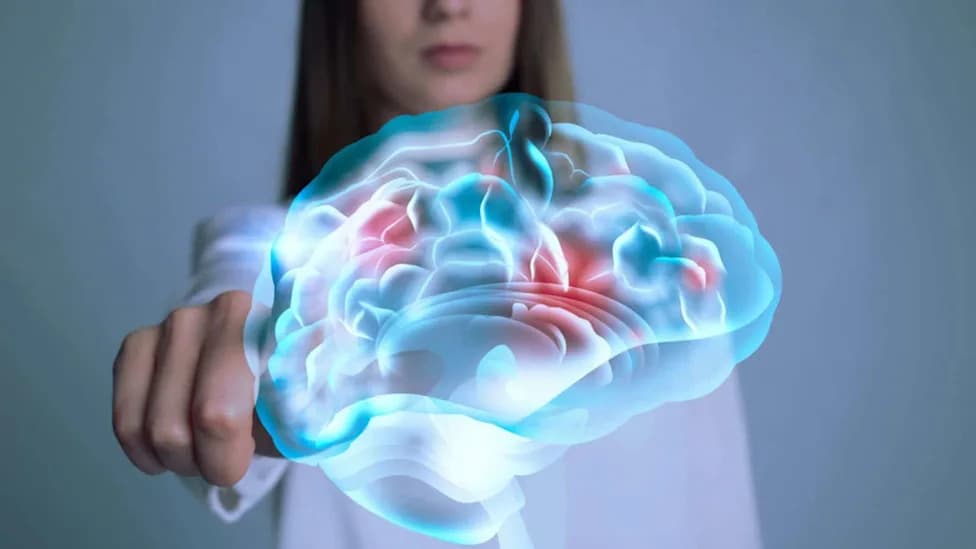
Scientists Found a Clue to How Anesthesia WorksSCIENCE PHOTO LIBRARY - Getty Images
Anesthesia has been medicine’s most reliable way to suspend consciousness for roughly 170 years, yet exactly how it silences awareness has remained unclear. Two new studies using complementary brain-imaging methods now map how brain rhythms reorganize as propofol drives people from wakefulness into unconsciousness—and reveal patterns that could improve monitoring and deepen our understanding of consciousness itself.
What the Studies Did
fMRI study (Frontiers in Computational Neuroscience): Researchers scanned 17 healthy adults with fMRI while they were gradually sedated with propofol through four defined states—awake, light sedation, deep sedation, and recovery. During each state, participants listened to a five-minute audio clip from the film Taken to probe auditory responsiveness. The team analyzed how spatial-temporal oscillatory modes (the brain’s eigenmodes) changed across states and trained a machine-learning model on those spatial patterns.
EEG study (Cell Reports Medicine): A separate group used high-resolution EEG to track electrical brain rhythms as subjects lost and regained consciousness under propofol. The EEG analysis focused on how low-frequency, large-scale network rhythms and specific oscillatory pathways (alpha, beta, gamma) behave during the transition into unconsciousness.
Key Findings
- Rather than acting as a simple on/off switch, anesthesia reorganizes brain oscillations. As consciousness declines, slow, large-scale low-frequency modes that span visual and somatomotor regions weaken, while faster, more local high-frequency activity increases in limbic (emotion and memory) areas.
- Auditory inputs continue to reach primary auditory cortex under sedation, but those signals fail to propagate to higher-order regions—consistent with fragmented, locally confined processing.
- The fMRI-derived spatial patterns of oscillatory change (eigenmodes) allowed a machine-learning classifier to identify a subject’s conscious state with about 72% accuracy using only spatial-index vectors—highlighting the diagnostic potential of these signatures.
- EEG results corroborated the fMRI findings by showing collapse of low-frequency network rhythms and an uncoupling of feedforward-feedback channels associated with alpha, beta, and gamma oscillations. As large-scale coordination breaks down, brain activity becomes locally synchronized and higher-order processing falters.
Why This Matters
Together the studies support a parsimonious view: conscious experience depends on widespread, low-frequency integration that broadcasts sensory information into higher-order cortex. When those broad rhythms collapse and activity becomes locally synchronized, the brain can detect stimuli but cannot integrate them into the complex processing needed for reportable awareness. That insight refines our theoretical picture of consciousness and points toward objective neural markers that could improve intraoperative monitoring of anesthesia depth.
Limitations And Next Steps
- Sample sizes were modest (particularly the fMRI study with 17 participants); results require replication in larger and more diverse cohorts.
- Both studies used propofol; different anesthetic agents may produce distinct patterns.
- Translating these findings into clinical monitoring tools will require robust, real-time metrics and validation across surgical populations and age groups.
Concluding Thought
These complementary fMRI and EEG studies move us closer to answering how anesthesia silences consciousness: not by globally turning the brain off, but by fracturing the large-scale rhythms that knit perception and cognition together. The discoveries open avenues for safer anesthesia management and for testing theories of consciousness with measurable neural signatures.
Help us improve.