Nikon is re‑reviewing the first‑place video from its Small World In Motion Competition after the microscopy community raised concerns that AI‑assisted post‑processing may have altered biological features. The winning entry, by Dr. Ning Xu of the National University of Singapore, shows abnormal airway cilia motion associated with primary ciliary dyskinesia; Xu says AI was used only to enhance visualization and that colors are false. Critics have called for release of the raw data and argue that AI 'hallucinations' could violate contest rules and scientific transparency. Nikon says Xu has provided technical documentation and will update the community after completing its review.
Nikon Re-Reviews Microscopy Video Winner After Concerns Over AI‑Assisted Post‑Processing
Nikon re-reviews video contest winner after complaints about AI use
Nikon is conducting a re‑review of the first‑place entry in this year’s Small World In Motion Competition after members of the microscopy community raised concerns that artificial intelligence was used to alter the winning video’s imagery.
Nikon Says Re‑Review Underway
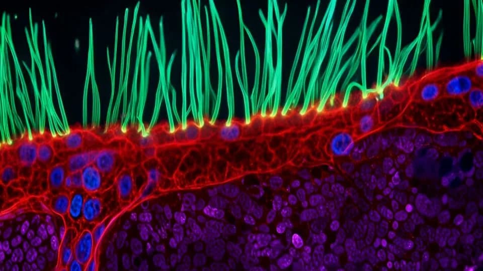
In a LinkedIn comment posted Sept. 26, Nikon said it is "carefully re‑reviewing" the first‑place video submitted by Dr. Ning Xu, a research fellow at the National University of Singapore. The short microscopic film depicts abnormal beating of airway cilia in a child with primary ciliary dyskinesia (PCD)—a rare genetic disorder that affects respiratory function—and Nikon's website notes that AI assisted the video’s post‑processing.
What Is At Issue
Contest rules for Nikon's Small World In Motion Competition prohibit AI‑generated videos. After the winning clip was posted on the Small World site and the contest's LinkedIn account, several microscopy professionals flagged aspects of the footage that they said appeared to be generated or significantly altered by AI tools.
'The point of image processing is to help us see (and quantify) structures we otherwise cannot resolve, not to hallucinate and make up things in your biological sample,' wrote Valentin Dunsing‑Eichenauer, a researcher at Der Simulierte Mensch in Berlin, in a comment on Nikon Small World’s post. He urged Xu to release raw data 'for the sake of transparency and fairness to the other competitors.'
Other commentators argued that post‑processing that invents plausible but fabricated biological features would violate the contest’s standards and could undermine scientific transparency and reproducibility.
Xu's Response
Dr. Xu replied in the LinkedIn comments, saying the team used AI‑assisted post‑processing to improve visual presentation and to distinguish features in grayscale super‑resolution images. He emphasized the colors in the video are false color and do not correspond to fluorescence channels that would identify specific cells or organelles. Xu wrote the rendered regions beneath the cilia were intended to enhance visualization rather than provide an anatomical reconstruction, and he said a detailed description of the optical system and a quantitative analysis of ciliary motion are being prepared for publication.
Nikon's Actions And Next Steps
Nikon said Xu is "respectfully complying" with the re‑review and has supplied detailed technical documentation describing his microscopy hardware, imaging protocols and processing workflow. Nikon added a disclaimer beneath the video noting AI‑assisted post‑processing, and said it intended to update the Small World community in the week of Sept. 28.
Judging Panel And Outreach
This year’s Nikon Small World in Motion judging panel included Joe Dragavon (Director, Advanced Light Microscopy Core, University of Colorado Boulder), Quinten Geldhof (technical designer, Museum of Science, Boston), Corey S. Powell (science writer and editor), and Meredith Sagolla (Director, Center for Advanced Light Microscopy and Electron Microscopy, Genentech). USA TODAY reached out to Nikon, Dr. Xu and the judges for comment; Nikon and Xu had not responded to USA TODAY’s inquiries as of Oct. 1.
Broader Context: AI, Image Processing And Scientific Standards
The controversy highlights a broader tension in scientific imaging: routine processing clarifies structures and enables measurement, but AI tools that plausibly synthesize or 'hallucinate' features blur the line between enhancement and fabrication. Critics say such alterations can undermine reproducibility, mislead viewers and create an unfair advantage in competitions governed by rules that restrict AI‑generated content. Supporters of careful, transparent use argue that advanced processing can improve accessibility and public engagement if methods are fully disclosed and raw data are made available.
Related: FAU 'Art of Science' Showcase
The Nikon coverage originally included a roundup of visual entries from Florida Atlantic University’s Division of Research 'Art of Science' contest. Highlights included an octopus 'cephie' captured by scientific SCUBA divers, candled images of a developing loggerhead sea turtle embryo, a high‑altitude balloon rupture photographed near 98,000 feet, deep‑space imaging of the Rosette Nebula compiled from 1,260 frames, confocal images of zebrafish radial glia, SEM images of coral skeleton microstructure, MRI montages of fruit cross‑sections, and other research images that illustrate how specialized imaging techniques turn scientific data into striking visual narratives.
As Nikon completes its re‑review, the case will be watched closely by microscopy professionals and imaging competitions as they wrestle with evolving standards around AI, image processing and scientific transparency.
Reporter: Greta Cross, USA TODAY
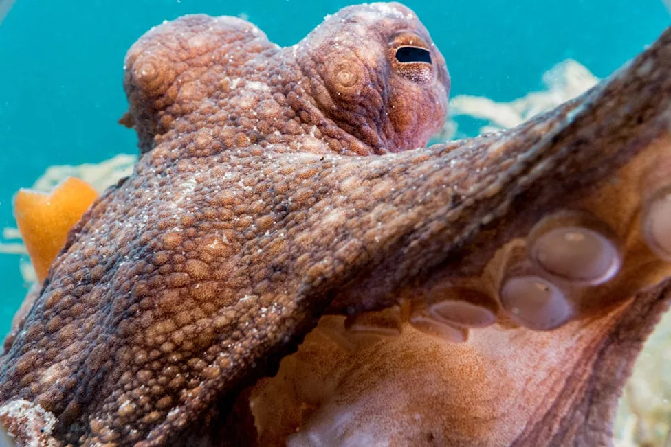
Florida Atlantic University’s Division of Research celebrates creativity with its annual “Art of Science” contest where research becomes stunning works of visual expression. Take a look at the mesmerizing images, starting with the competition's first place winner ...FIRST PLACE: SELFIE ... CEPHIE.Octopuses have eight flexible appendages that can bend, shorten, elongate and twist in all directions due to their extensive nervous system and the complex arrangement of their arm musculature, which lack bones. Scientific SCUBA divers held their cameras close as the octopus tried to take a “cephie,” or octopus selfie.(Photography by Chelsea Bennice,Ph.D.,postdoctoral fellow,Charles E. Schmidt College of Science)
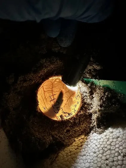
SECOND PLACE: SEA TURTLE BEGINNING.Through a method called candling, a flashlight is held up against an egg in the dark to visualize embryonic development. The flipper outline of a developing loggerhead sea turtle (Caretta caretta) embryo is visible within its egg, along with the intricate network of extraembryonic blood vessels that sustain growth.(Photography by Gabriella Carvajal,doctoral student,Charles E. Schmidt College of Science,Mentor: Jeanette Wyneken,Ph.D.)
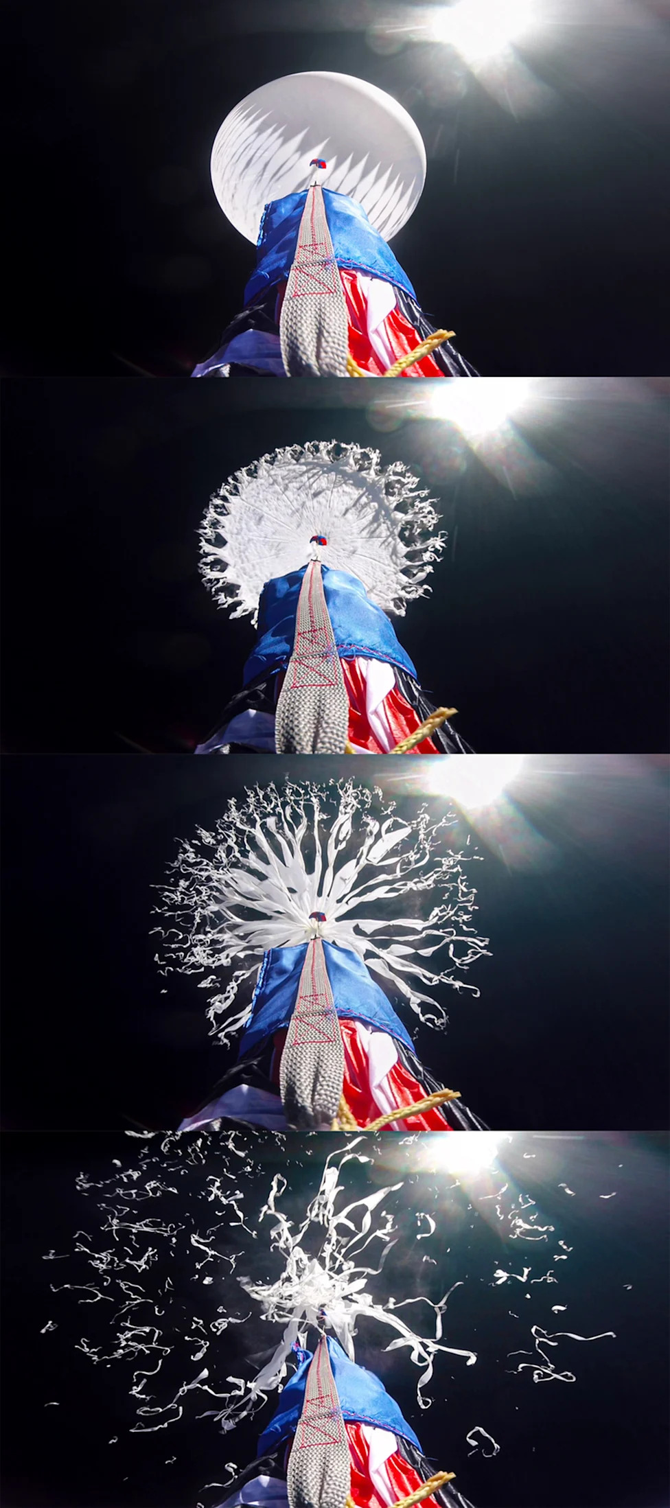
THIRD PLACE: JOURNEY TO SPACE.This image sequence was captured at about 98,000 feet during the Aerospace Experimental Association’s Mission 3, a high-altitude balloon launch conducted in fall 2024 at Florida Atlantic University. It shows the precise instant when the balloon ruptured at the edge of space, marking the transition from ascent to descent. The photograph was taken by a GoPro mounted at the top of the payload, facing upward.(Photography by Sky Rueff ,undergraduate student,College of Engineering and Computer Science Mentor: Oscar Curet,Ph.D.)
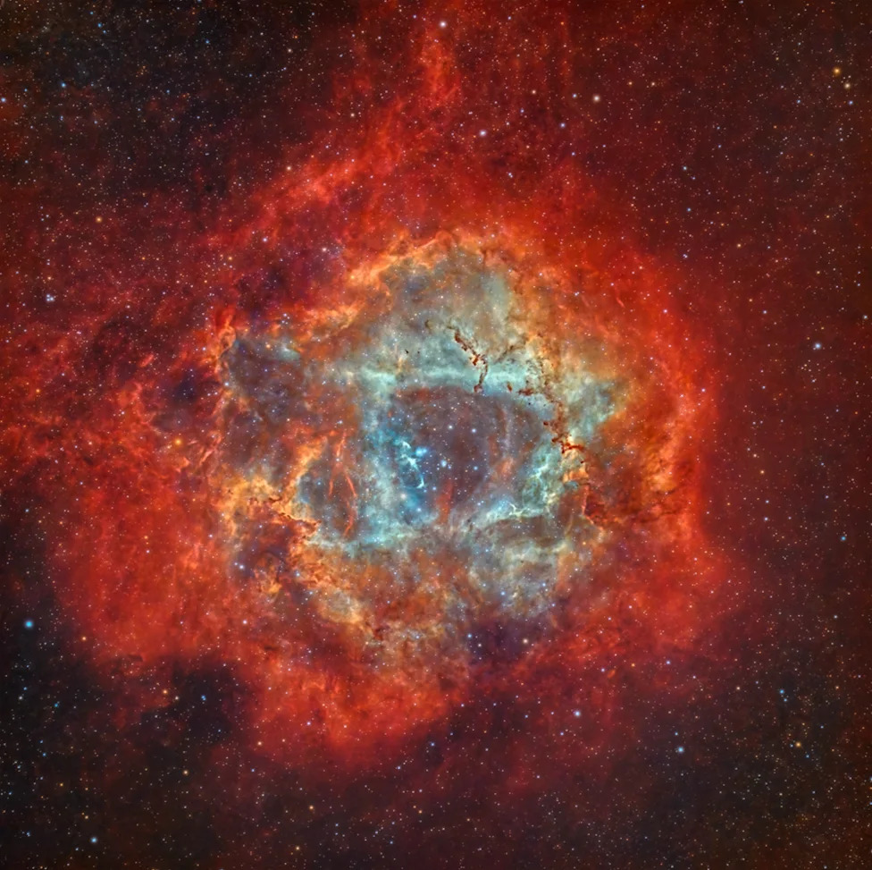
FACULTY IN THE FIELD: ROSETTE NEBULA.This shot is comprised of 1260 pictures totaling 10.5 hours of total imaging time. The Rosette Nebula is located approximately 5,200 light-years from Earth. It is a large cloud of ionized hydrogen gas. The vibrant red colors seen in images of the Rosette Nebula are primarily due to the emission of light from these hydrogen atoms. The nebula is estimated to have a diameter of about 130 light-years.(Photography by Rob Gross,Ph.D.,instructor Charles E. Schmidt College of Science)
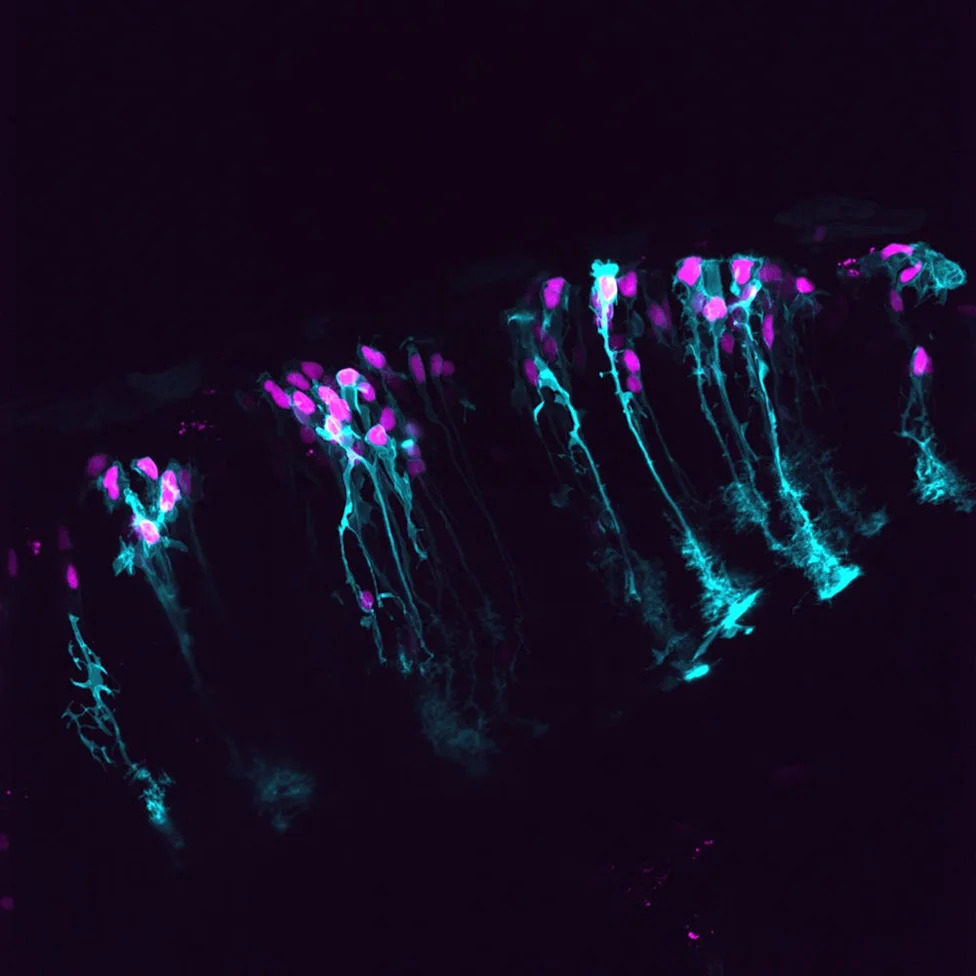
FACULTY IN THE LAB: RADIANT ARCHITECTS.This confocal microscopy image captures radial glial cells in the zebrafish central nervous system, highlighting their unique morphology and critical role in neural development.(Photography by Laura Fontenas,Ph.D.,assistant professor,Charles E. Schmidt College of Science)
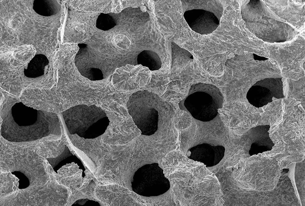
STUDENT IN THE LAB: SKELETAL SECRETS.Coral reefs are crucial players for their marine ecosystems by protecting coastal areas, promoting biodiversity and providing a plethora of ecological benefits for cohabitating wildlife. This is a scanning electron microscope (SEM) image of P. astreoides at 120x magnification depicting the morphology and microstructures of its skeleton.(Photography by Alejandra Coronel-Zegarra,doctoral student,Charles E. Schmidt College of Science,Mentor: Vivian Merk,Ph.D.)
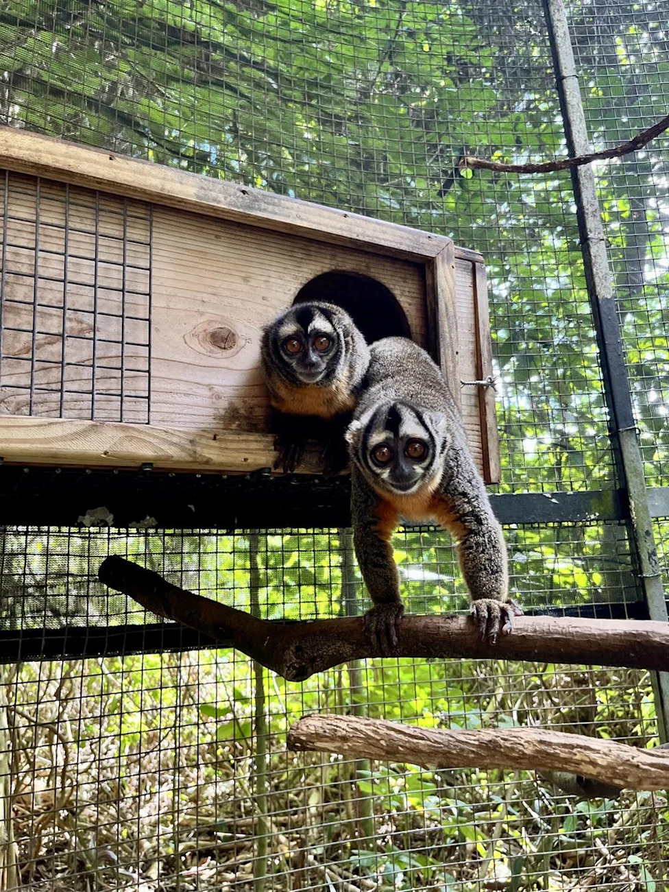
STUDENT IN THE FIELD: TWO TO TANGO.Ever tried an activity like kayaking with a partner, only to discover that coordinating paddling is harder than it looks, and end up stuck in some mangroves?(Photography by Aaron Mencia,graduate student,Charles E. Schmidt College of Science,Mentor: Kate Detwiler,Ph.D.)
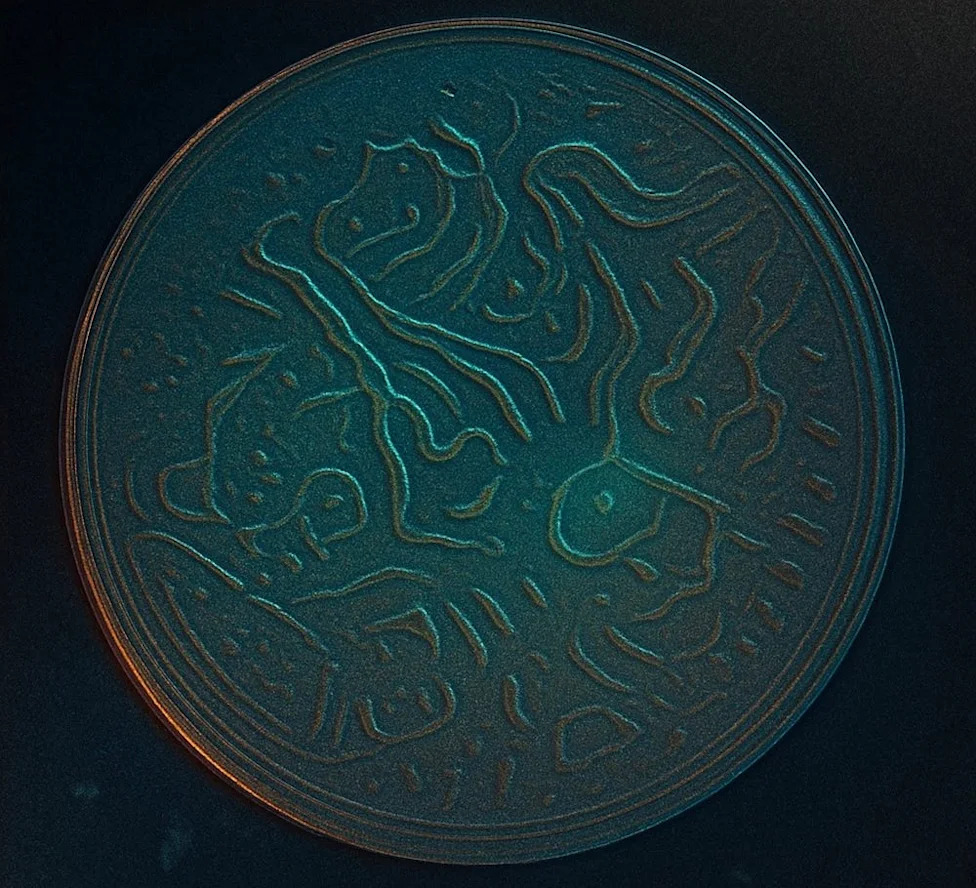
TOP POSTDOC: SURVIVAL.Here, the C. elegans nematodes were left searching for more food, and instead of hiding their need, they etched their hunger into the agar, leaving behind an abstract pattern. What looks like art is actually survival - a biological request written in unexpected lines.(Photography by Hussam Alshareef,Ph.D.,postdoctoral fellow,Stiles-Nicholson Brain Institute)
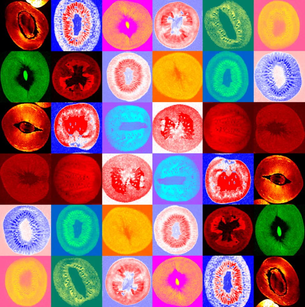
TOP STAFF: ORCHARD ARCHITECTURE.This montage presents fruit in an entirely unique way - through the lens of magnetic resonance imaging (MRI). By capturing cross-sections with exquisite clarity and without invasive techniques, MRI reveals the hidden architecture of seeds, rinds and pulps in remarkable detail. What appears at first like abstract art is, in fact, the internal world of familiar produce, transformed into patterns of symmetry, texture and contrast.(Photography by Refaat Gabr,Ph.D.,staff ,Division of Research)
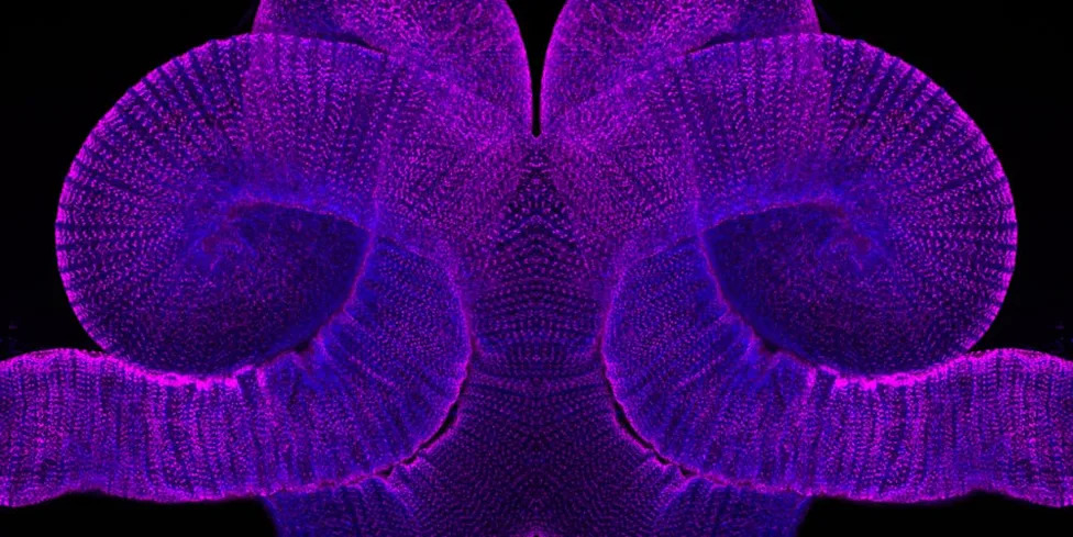
RESEARCH EXCELLENCE: FACE OF DIGESTION.The picture shows a double-labeled of the fruit fly gut. Animals were genetically modified to add a fluorescent-tag (blue color) to an endogenous protein, which has been linked to metabolic disorders such as diabetes and obesity. This allows for visualization and study expression differences of the protein in response to different diets, such as high sugar, high protein or no food. The specimen was also immunohistochemically labeled with antibodies against a structural gut protein (magenta color) to visualize the gut structure. Confocal microscopy was used to acquire images of each fluorescent label.(Photography by Emily Arbesfeld, undergraduate student, Harriet L. Wilkes Honors College, Mentor: Tanja Godenschwege, Ph.D.)
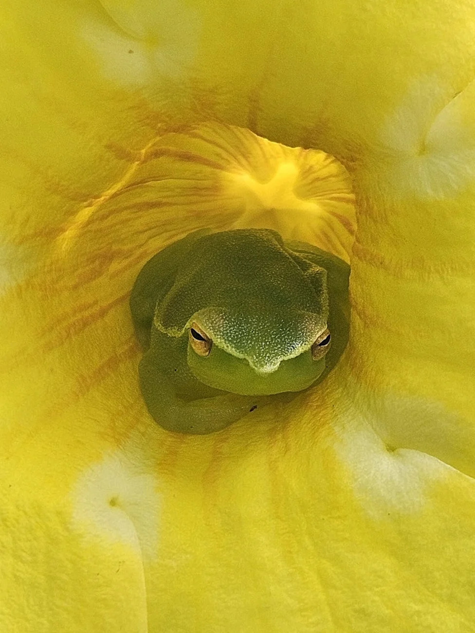
RESEARCH EXCELLENCE: HATCHET.A type of lime treefrog, the picture shows the distinctive "hatchet" face of Sphaenorhynchus lacteus. The picture was captured near the bank of the only known boiling river in the world, called Mayantuyacu, in a remote area of the central Peruvian Amazon.(Photography by Yash Bhagwanji, Ph.D., associate professor, College of Education)
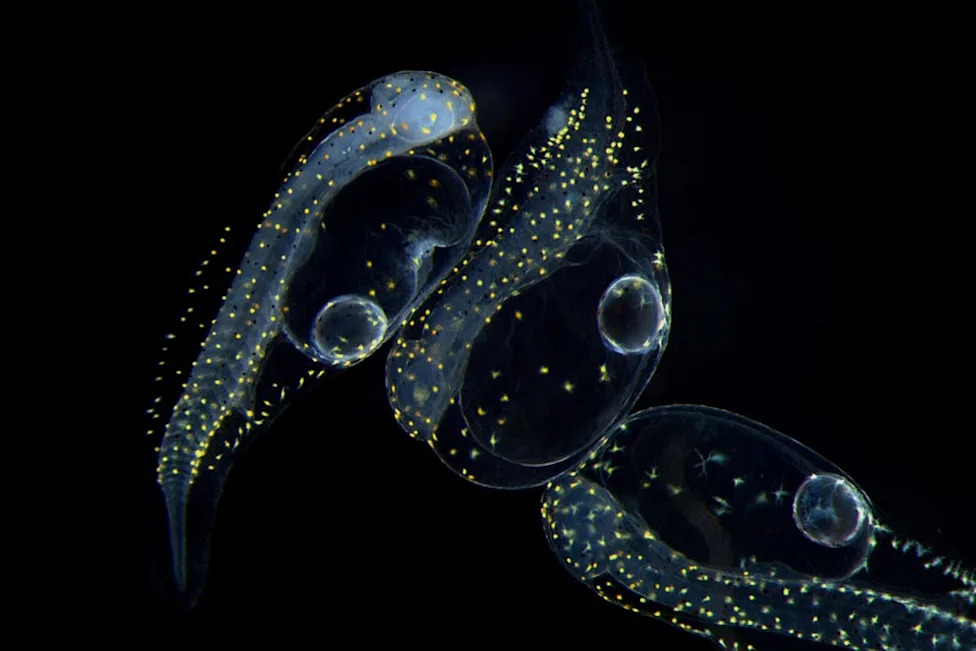
RESEARCH EXCELLENCE: TRES LECHES.These are newly hatched Florida pompano (Trachinotus carolinus) larvae reared at the Florida Atlantic University Harbor Branch Oceanographic Institute. At just 0 days post-hatch, they have only recently emerged from their eggs and represent the earliest stage of development for this coastal species.(Photography by Li Sun Chin, doctoral student, Charles E. Schmidt College of Science and FAU Harbor Branch Oceanographic Institute, Mentor: Paul Wills, Ph.D.)
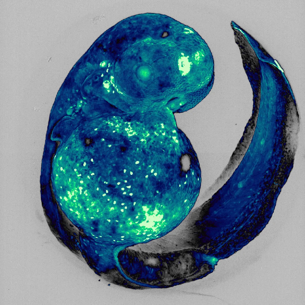
RESEARCH EXCELLENCE: ORIGIN STORY.This image captures a developing zebrafish embryo, a powerful model for exploring how the nervous system takes shape. The image has been falsely colored for artistic effect, highlighting the intricate beauty of developing structures that guide our understanding of both healthy and disordered development.(Photography by Tessa Dallo, doctoral student, Charles E. Schmidt College of Science and Stiles-Nicholson Brain Institute, Mentor: Laura Fontenas, Ph.D.)
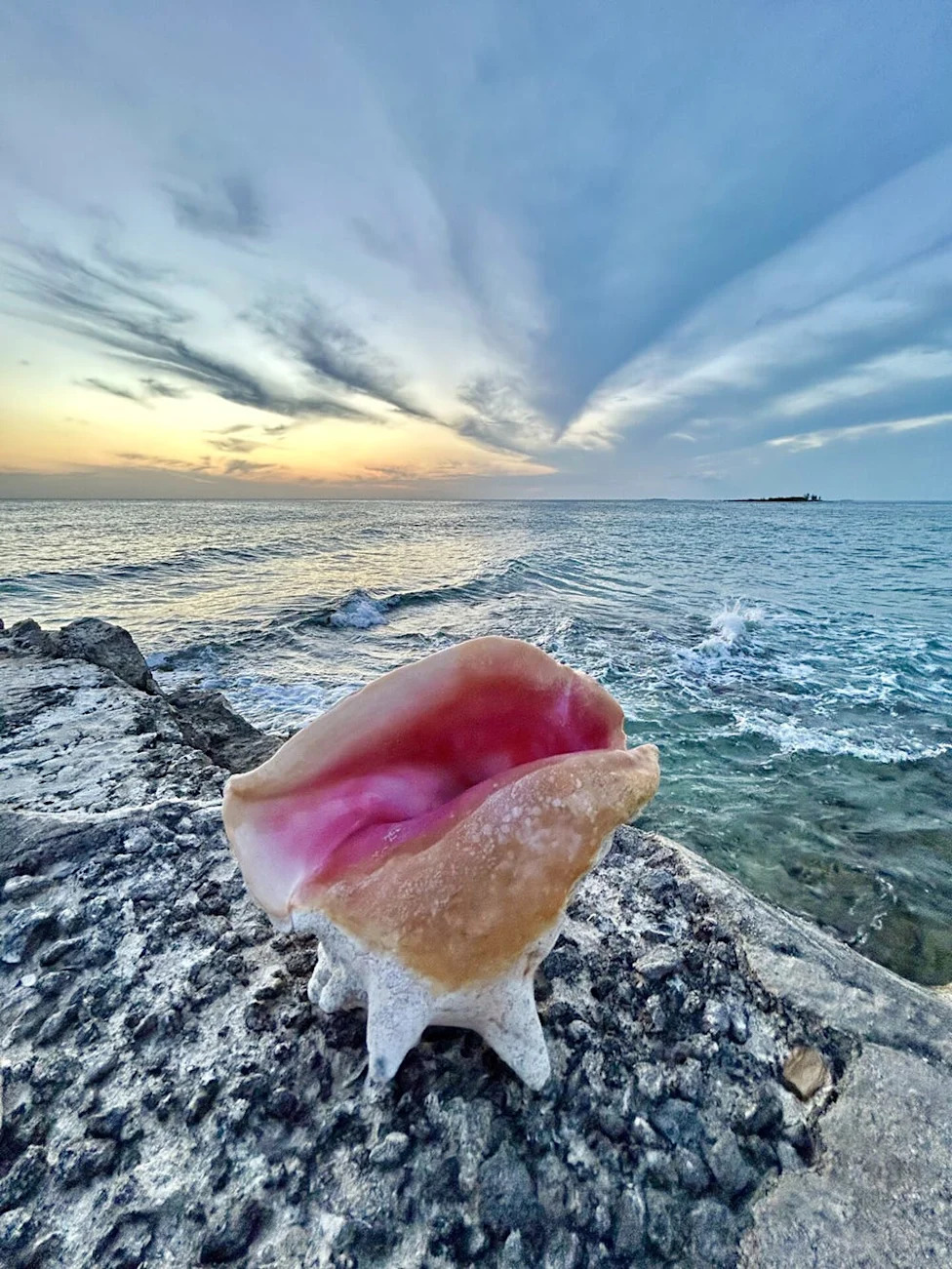
RESEARCH EXCELLENCE: CONCH SUNSET.The FAU Queen Conch Lab has a partnership project with Blue Action Lab on Grand Bahama Island. The Mobile Labs are used to raise queen conch larvae to juvenile stage for conservation, restoration and education. The queen conch is important for the culture, nutrition, livelihood and coastal seas of countless communities in The Bahamas.(Photography by Megan Davis, Ph.D., research professor, Harbor Branch Oceanographic Institute)
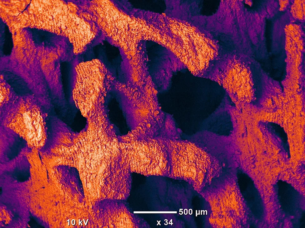
RESEARCH EXCELLENCE: WHERE BONE MEETS GOLD.False-colored SEM image of 3D-printed beta-tricalcium phosphate (β-TCP) for bone regeneration. Bone has an incredible, but limited, ability to repair itself. Traditionally, the gold standard treatment is bone transplantation from another healthy site in the same patient. However, this involves a second surgery, increasing the risk of complications and treatment costs. This image shows a synthetic alternative: 3D-printed β-TCP, a ceramic material with a calcium-phosphate composition similar to natural bone. The structure has a multi-scale interconnected porous architecture (350—1500µm) and fine supporting struts (230—550µm). The manufacturing of such complex geometry is only achievable through 3D printing, resulting in an implant that is both beautiful and functional. Over time, the β-TCP biodegrades and is naturally replaced by the patient’s own bone.(Photography by Ahmed Eltawila, doctoral student, College of Engineering and Computer Science, Mentor: Yunqing (Kevin) Kang, Ph.D.)
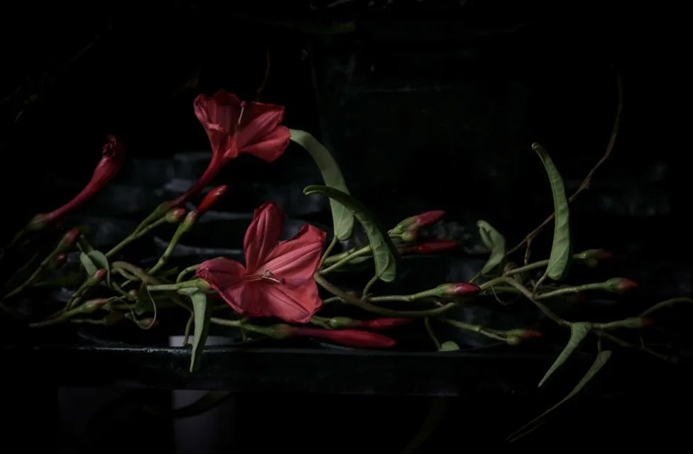
RESEARCH EXCELLENCE: MAN-IN-THE-GROUND VINE.Man-in-the-ground vine (Ipomoea microdactyla) is state-listed as endangered. The photographer is researching flora and fauna native to Florida, with an emphasis on at-risk and endangered species. The subjects of the portraits are photographed within their natural habitats or at local conservation organizations. They range from a rare deep red morning glory found primarily in the pine rocklands of Miami-Dade County (man in the ground/Ipomoea microdactyla) to the loggerhead sea turtle (Caretta caretta), which faces multiple threats, including habitat loss, light pollution and incidental capture by fisheries. The project merges artistic observation with ecological inquiry to reflect Florida’s biodiversity.(Photography by Sharon Hart, MFA, professor, Dorothy F. Schmidt College of Arts and Letters)
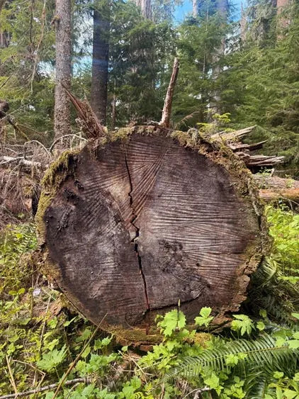
RESEARCH EXCELLENCE: CRACKS THAT MEND.This image of a fallen tree reveals the story of time, struggle and survival. Each ring marks a year of growth, while the cracks running through the wood tell of storms weathered and pressures endured. Despite the breaks, the tree remains whole and offers shelter to new plants and moss along its edges. In many ways, this mirrors the human journey through mental health. Life leaves its marks, some visible and some hidden, but resilience is found in how we continue to grow around the fractures. Healing does not erase the cracks; it transforms them into part of our strength and story.(Photography by Maria Mejia, professor, Charles E. Schmidt College of Medicine)
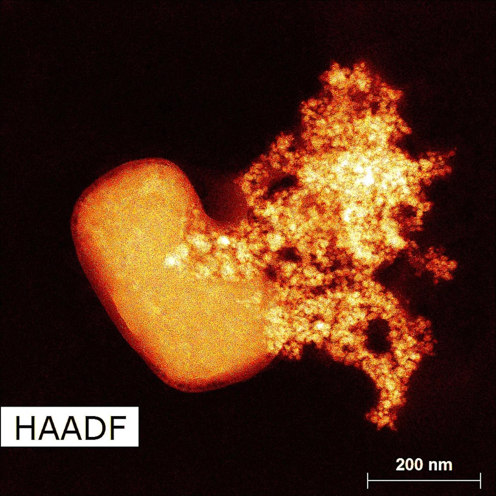
RESEARCH EXCELLENCE: METALLIC BLOOM.This image was captured using specialized imaging called High-Angle Annular Dark Field with a specific type of microscope called scanning transmission electron microscopy. It uses electrons instead of light to reveal structures far smaller than what regular microscopes can detect. The bright cluster shows nickel particles formed by heating chemicals to 170°C in a polyol liquid called ethylene glycol. The polyol acts like a chemical cooking medium, helping control how the nickel particles form and grow. Though the particles didn’t grow on the salt crystal, the surface beneath them looks like one, creating the illusion of a metallic bloom. Each particle is merely a billionth of a meter wide, several times smaller than a grain of sand, yet together they form a branching pattern shaped by heat and chemistry.(Photography by Gladel Saintilme, graduate student, Graduate College, Mentor: Vivian Merk, Ph.D.)
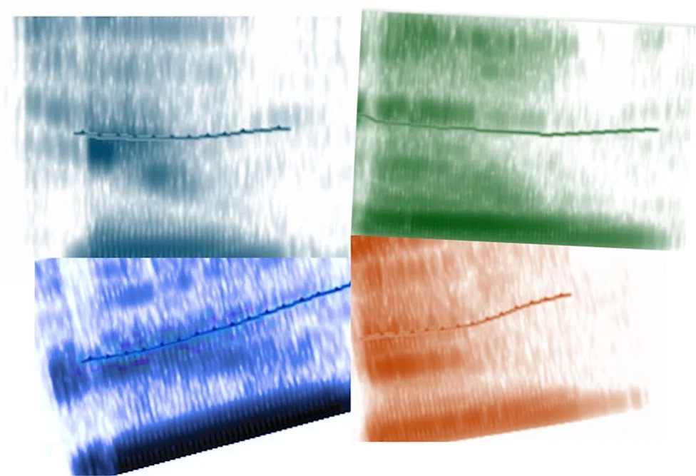
RESEARCH EXCELLENCE: SONIC PORTRAIT.This artwork traces the acquisition of the Danish vowel /y/, as in hygge. Through repeated attempts, the speaker’s pronunciation shifts — each spectrogram capturing a moment of articulation in transition. These four images are voiceprints: acoustic and visual records of a phoneme being learned. Subtle changes in formant structure reflect the fine motor adjustments of speech. The data were generated and analyzed using Praat, an open-source tool for phonetic research. Together, the images form a sonic portrait of a vowel, a study in adaptation, where speech and language takes shape through repetition and time.(Photography by Anne L. van Zelst, Ph.D., assistant professor, College of Education)
Help us improve.