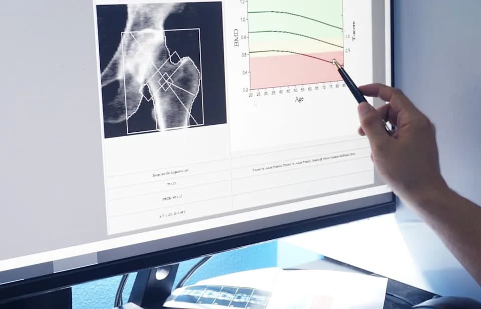
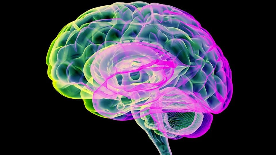
A large multimodal study of more than 18,000 UK Biobank participants finds that where you store fat—not just how much you weigh—strongly correlates with differences in brain aging. Visceral fat (around organs) was uniquely associated with white-matter damage, a change linked to vascular cognitive impairment and higher dementia risk. Researchers propose chronic inflammation and metabolic disruption as likely mechanisms but stress the study is observational and cannot prove causation. Because visceral fat is modifiable, reducing it through lifestyle or clinical measures could offer new paths for prevention pending further trials.
Where You Store Body Fat Predicts Brain Aging — Large Study Links Visceral Fat To White-Matter Damage
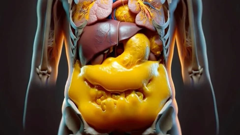
(Mykola Chaika/Creatas Video+/Getty Images Plus)
People with the same body weight or BMI can carry very different risks to brain health depending on where they store fat, according to a large new analysis of UK Biobank data. Researchers at The Hong Kong Polytechnic University (PolyU) mapped regional adiposity against brain scans and cognitive tests for more than 18,000 participants and report distinct brain changes associated with fat in different body regions.

YouTube Thumbnail
Key Findings
The study—published in Nature Mental Health—is the largest multimodal analysis to systematically examine how the location of body fat relates to brain structure, connectivity and cognitive performance. The authors found that:
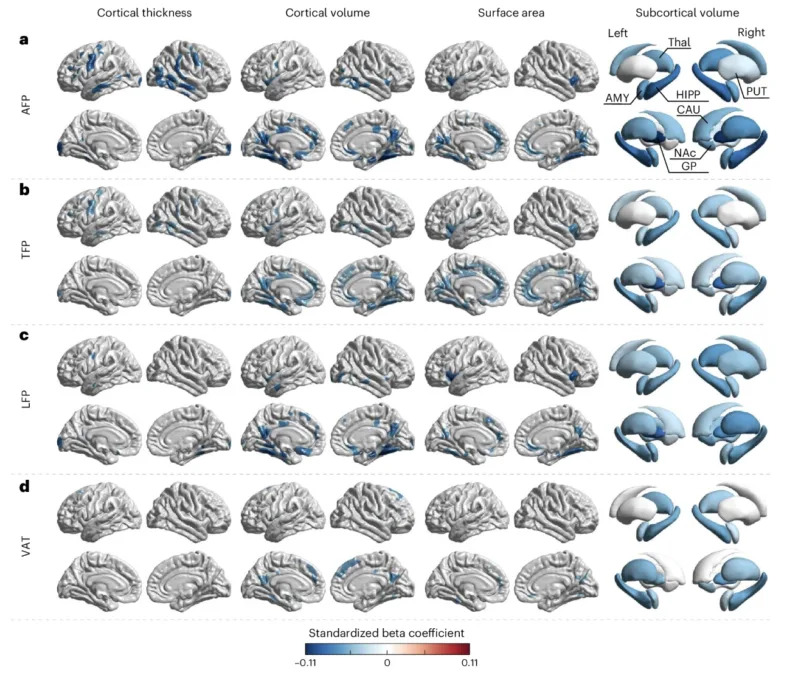
The relationships between specific body fat storage and brain morphology. (Zhang et al.,Nat. Ment. Health., 2025)
- Visceral adipose tissue (VAT), the internal fat that surrounds organs, was uniquely associated with damage to the brain’s white matter—the network of nerve fibres that transmits signals between brain regions.
- Fat percentages in the arms, legs and trunk mapped to different, region-specific patterns of brain change (affecting systems involved in movement, emotion, memory and brainstem functions) that were distinct from the pattern linked to visceral fat.
Why It Matters
White-matter deterioration is closely tied to the biology of vascular cognitive impairment and cerebral small vessel disease—conditions that increase the risk of dementia. The authors argue that visceral fat may promote chronic, low-grade systemic inflammation and metabolic dysregulation (including insulin and glucose pathways) that spill over to the brain and drive neuroinflammation and white-matter injury.
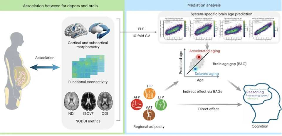
A graphical summary of the study. Left: associations between regional adiposity and brain morphology, functional connectivity and white-matter microstructure. Right: construction of a brain age prediction model. (Zhang et al.,Nat. Ment. Health., 2025)
“Fat in different parts of the body maps out entirely different trajectories of change in the brain, which cannot be detected by body weight or BMI alone,” says PolyU neuroinformatician Anqi Qiu. “Our analytical model clearly demonstrates how fat in different regions causes differential damage to brain systems.”
Methods Brief
The team analyzed imaging-derived measures of regional fat (arm, leg, trunk, and visceral fat), brain morphology, functional connectivity and white-matter microstructure, alongside cognitive test scores, in more than 18,000 UK Biobank participants. They also constructed a brain-age prediction model to estimate associations between regional adiposity and apparent brain aging (Zhang et al., Nat. Ment. Health., 2025).

Subscribe to ScienceAlert's free fact-checked newsletter
Limitations and What We Don’t Yet Know
- The analysis is observational and cannot establish causation—other factors could contribute to both fat distribution and brain health.
- Biological mechanisms remain hypothetical: inflammation and metabolic pathways are plausible, but direct causal links need validation in longitudinal and interventional studies.
- The study population (UK Biobank) is large but not fully representative of all ethnic and age groups, so results may not generalize to every population.
Practical Takeaways
Regional adiposity is a potentially modifiable risk factor. Unlike overall BMI, where two people can look the same, visceral fat appears to carry disproportionate risk for brain aging. The authors suggest that targeted lifestyle or clinical interventions that reduce visceral fat—through diet, physical activity, sleep, and metabolic management—could provide new avenues for preventing or delaying neurodegenerative disease, but randomized trials are needed.
Bottom line: Where you store fat matters. Visceral fat around internal organs is the type most clearly linked to brain white-matter damage and accelerated brain aging in this large multimodal study, but further research is needed to confirm a causal role and to test interventions.
Reference: Zhang et al., Nature Mental Health, 2025. Data source: UK Biobank imaging and cognitive datasets. Quotes and interpretation from PolyU researchers.
Help us improve.